Full Text (PDF)
Indian Journal of Forensic Medicine and Pathology 18(2):p 131-136, Jan-June 2025. | DOI: https://doi.org/10.21088/ijfmp.0974.3383.18225.7
Case Report
Unravelling the Secrets of Age Estimation: A Look at the Science Behind the Bone Analysis
Roopam Mourya, Ramkrishna Mishra, Surendra Kumar Pandey, Devashish Verma
Author Information
Licence:
Attribution-Non-commercial 4.0 International (CC BY-NC 4.0)This license enables reusers to distribute, remix, adapt, and build upon the material in any medium or format for noncommercial purposes only, and only so long as attribution is given to the creator.
Indian Journal of Forensic Medicine and Pathology 18(2):p 131-136, Jan-June 2025. | DOI: https://doi.org/10.21088/ijfmp.0974.3383.18225.7
How Cite This Article:
Mourya R, Mishra R, Pandey SK, et al. Unravelling the Secrets of Age Estimation: A Look at the Science Behind the Bone Analysis. Indian J Forensic Med Pathol. 2025;18(2):131-136.Timeline
Received : March 15, 2025
Accepted : May 30, 2025
Published : June 20, 2025
Abstract
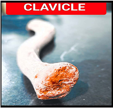
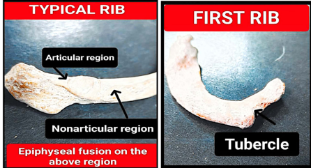
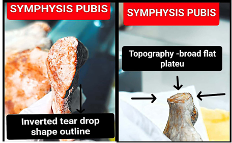
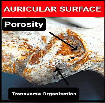
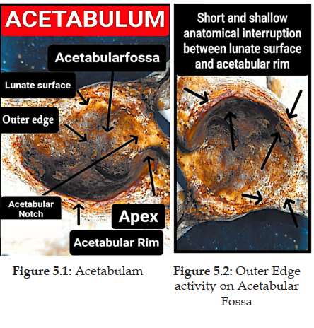
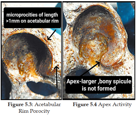
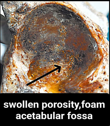
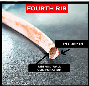
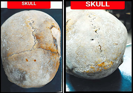
Determining the age of unidentified human skeletal remains is one of the objectives of forensic identification. Age is estimated using the articular surface of the ilium, pubic symphysis, acetabulum, clavicle, skull, and sternum. In November 2020, the body of an unknown 35-year-old man was found in a black plastic bag and transported to the Varanasi postmortem house for examination. Age can be inferred from changes in bone morphology as people age. The age had been estimated by the police. Bone morphology is the most accurate indicator of age, especially in a medicolegal setting. The deceased’s age is determined to be between 40 and 45 years old after a postmortem. For this middle age group estimation of age, most reliable morphological changes occur in acetabulum, pubic symphysis and the 4th rib.
References
- 1. Moraitis K, Zorba E, Eliopoulos C, et al. A test of the revised auricular surface aging method on a modern European population. J Forensic Sci. 2014;59(1):188-94.
- 2. Cerezo RJI, Hernåndez EPO. Estimating age at death using the sternal end of the fourth ribs from Mexican males. Forensic Sci Int. 2014;236:196.1-6.
- 3. Loth SR, Iscan MY. Morphological age estimation. In: Seigal J, Knupfer G, editors. Encyclopedia of forensic sciences. [place unknown]: [publisher unknown]; 2010.
- 4. Pickering R, Bachman D. The use of forensic anthropology. [place unknown]: [publisher unknown]; 2009. p. 1.
- 5. Anderson MF, Anderson DT, Wescott DJ. Estimation of adult skeletal age-at-death using the Sugeno fuzzy integral. Am J Phys Anthropol. 2010;142(1):30-41.
- 6. Passalacqua NV. Forensic age-at-death estimation from the human sacrum. J Forensic Sci. 2009;54(2):255-62.
- 7. Black S, Scheuer B. Age changes in the clavicle: from the early neonatal period to skeletal maturity. Int J Osteoarchaeol. 1996;6:425-43.
- 8. Meijerman L, Maat GJR, Schulz R, Schmeling A. Variables affecting the probability of complete fusion of the medial clavicular epiphysis. Int J Leg Med. 2007;121:463-8.
- 9. Scheuer L, Black S. Illustrations by Angela Christie, Developmental Juvenile Osteology. [place unknown]: Elsevier Academic Press; 2000. p. 251. (The pectoral girdle, the clavicle).
- 10. Singh J, Chavali KH. Age estimation from clavicular epiphyseal union sequencing in a Northwest Indian population of the Chandigarh region. J Forensic Leg Med. 2011;18.
- 11. Scheuer L, Black S. Illustrations by Angela Christie, Developmental Juvenile Osteology. [place unknown]: Elsevier Academic Press; 2000. p. 231-42. (The thorax, the ribs and costal cartilages).
- 12. Aggrawal A. Textbook of forensic medicine and toxicology. 2nd ed. [place unknown]: Avichal Publication; 2021. Identification, sternum; p. 81-2.
- 13. Truesdell J. The composite method: a novel, continuum-based approach to estimating age from the female pubic symphysis with particular relevance to mature adults. Forensic Sci. 2023;3(1):94-119. doi:10.3390/forensicsci3010009.
- 14. Priya E. Methods of skeletal age estimation used by forensic anthropologists in adults: a review. Forensic Res Criminol Int J [Internet]. 2017 [cited 2025 Sep 18];4(2):41-51. Available from: https://dx.doi.org/10.15406/frcij.2017.04.00104.
- 15. Martha San Millan, Carme Rissech, et al. New approach to age estimation of male and female adult skeletons based on the morphological characteristics of the acetabulum. Int J Legal Med [Internet]. 2017 Mar [cited 2025 Sep 18];131(2):501-25. Available from: https://dx.doi.org/10.1007/s00414-016-1406-4.
- 16. Meena MC, Rani Y. Age estimation from the IV rib by the components method in Indian males. Aust J Forensic Sci [Internet]. 2014 [cited 2025 Sep 18];2014 Mar 20:463-70. Available from: [URL].
- 17. Aggrawal A. Textbook of forensic medicine and toxicology. 2nd ed. [place unknown]: Avichal Publication; 2021. Identification, Ossification center during IU life; p. 89.
Data Sharing Statement
There are no additional data available.
Funding
This research received no funding.
Author Contributions
All authors contributed significantly to the work and approve its publication.
Ethics Declaration
This article does not involve any human or animal subjects, and therefore does not require ethics approval.
Acknowledgements
Information not provide.
Conflicts of Interest
The authors report no conflicts of interest in this work.
About this article
Cite this article
Mourya R, Mishra R, Pandey SK, et al. Unravelling the Secrets of Age Estimation: A Look at the Science Behind the Bone Analysis. Indian J Forensic Med Pathol. 2025;18(2):131-136.
Licence:
Attribution-Non-commercial 4.0 International (CC BY-NC 4.0)This license enables reusers to distribute, remix, adapt, and build upon the material in any medium or format for noncommercial purposes only, and only so long as attribution is given to the creator.
| Received | Accepted | Published |
|---|---|---|
| March 15, 2025 | May 30, 2025 | June 20, 2025 |
DOI: https://doi.org/10.21088/ijfmp.0974.3383.18225.7
Keywords
Age estimationSkeletal remainsBone morphologyOssificationPorocityEpiphysisSearch for Similar Articles
Similar Articles
- Sudden Death in a 7-year-old Child Due to Neoplasm: A Case Report
- Bites of Burden: Snake Bite Envenoming in India, Regional Venom Differences, Tr...
- Forensic View of Suicide in Healthcare Workers
- Tracing Identity from Teeth to Technology: Artificial Intelligence in Forensic...
- Chemical Constituents in Latent Fingermarks as Indicators of Lifestyle using Gas...
Article Level Metrics
Last UpdatedWednesday 17 June 2026, 23:17:45 (IST)
7105
Accesses
21
2030
00
Citations
NA
NA
NA
Download citation
Article Keywords
Keyword Highlighting
Highlight selected keywords in the article text.
Timeline
| Received | March 15, 2025 |
| Accepted | May 30, 2025 |
| Published | June 20, 2025 |
licence
Attribution-Non-commercial 4.0 International (CC BY-NC 4.0)
This license enables reusers to distribute, remix, adapt, and build upon the material in any medium or format for noncommercial purposes only, and only so long as attribution is given to the creator.