Full Text (PDF)
Indian Journal of Anatomy 12(1):p 23-29, January-March 2023. | DOI: https://doi.org/10.21088/ija.2320.0022.12123.2
Original Article
Study of Anatomical Variants of the Temporal Bone Pen Process during Eagle’s Syndrome
Mar Ndeye Bigué, Loum Birame, Niang Ibrahima, Yacouba Garba Karim, Deme Hamidou, Akpo Géraud Léra, Diop Mamadou, Diallo Bay Karim, Ndiaye Assane
Author Information
Licence:
Attribution-Non-commercial 4.0 International (CC BY-NC 4.0)This license enables reusers to distribute, remix, adapt, and build upon the material in any medium or format for noncommercial purposes only, and only so long as attribution is given to the creator.
Indian Journal of Anatomy 12(1):p 23-29, January-March 2023. | DOI: https://doi.org/10.21088/ija.2320.0022.12123.2
How Cite This Article:
Mar NB, Loum B, Niang I, et al. Study of anatomical variants of the temporal bone pen process during Eagle’s syndrome. Indian J Anat. 2023;12(1):23–9.Timeline
Received : October 21, 2022
Accepted : November 24, 2022
Published : December 30, 2022
Abstract
Anatomical variations of the pen process (PS) and ossification of the pen-hyoid ligament constitute Eagle syndrome. This is a rare syndrome, often bilateral, that summarizes the clinical manifestations due to an excessive length of the pen process that is greater than 25mm. Other important morphometric and topographical variations are associated with this anomaly.
Taking these anatomical data into account is essential for the etiopathogenic analysis and surgical treatment of Eagle syndrome. The purpose of this work was to investigate anatomical variations of the pen process during Eagle syndrome.
Material and method: This was a retrospective study over a period of 07 months from December 2020 to June 2021 inclusive. It concerned 32 patients, received in the radiology and medical imaging department of the National Pikine Hospital for suspicion of Eagle syndrome confirmed by scanner without and with injection. Patients who had a normal scanner were not included in the study. We used SPSS 24.0 and Excel 2016 to analyze the data.
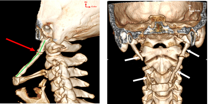
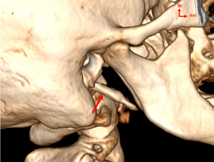
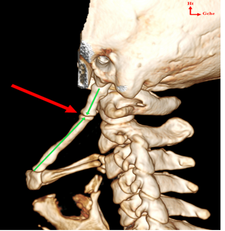
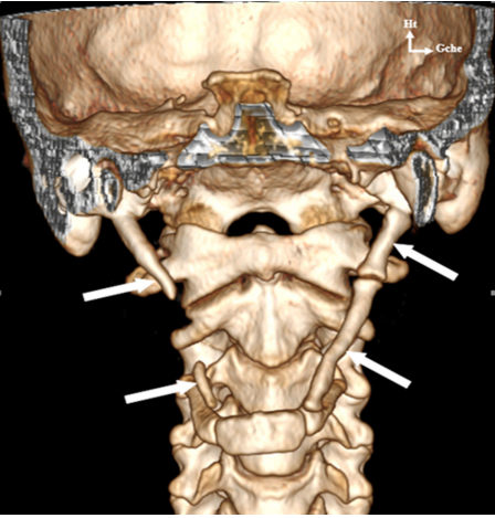
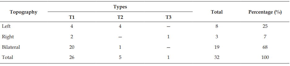
Results: 24 women (75%) and 8 men (25%), a sex ratio of 0.33. The average age of patients was 39 years, with a standard deviation of 12 years, and extremes of 21 to 71 years. The age range of 30-40 was 34% and 41-50 was 33%. Examination found pharyngeal foreign body sensation in 65% of cases, dysphagia in 67% of cases, headache in 71% of cases, and cervical neuralgia. Brachial in 95% of cases. At CT, the average length of all pen processes was 39.2 mm (extremes: 21-76 mm). The elongation of the pen process was bilateral in 68%, unilateral right in 7% of cases, and unilateral left in 25% of cases. Morphologically, the PS consisted of a single long fragment in 78% of cases (Langlais types 1), or two fragments articulated either by fibrous tissue or by a pseudo articulation with an angular shape in 19% of cases (Langlais types 2), or several fragments (3% Langlais types 3).
Conclusion: Eagle syndrome is a radio anatomical entity with several variants whose knowledge is essential to its management.
References
- 1. Eagle WW. Elongated styloid process: report of two cases. Arch Otolaryngol. 1937;25:584–7.
- 2. Balde D, Do Santos ZA, Ndiaye C, Dione A, Diallo BK, Adjibabi W, et al. Intra oral versus external approach in the surgical management of Eagle’s syndrome. Int J Otorhinolaryngol Head Neck Surg. 2019;5(1):9–14.
- 3. El Hassane. Apport de la tomodensitometrie dans le diagnostic du syndrome d’Eagle au Centre Hospitalier Regional Heinrich Lübke de Diourbel: à propos de 30 cas [Specialty Thesis]. Dakar: Université Cheikh Anta Diop de Dakar; 2019. Thesis No. 313. French.
- 4. Lengele B, Dhem A. Microradiographic and histological study of the styloid process of the temporal bone. Acta Anat (Basel). 1989;135(3):193–9.
- 5. Raser JM, Mullen MT, Kasner SE, Cucchiara BL, Messe SR. Cervical carotid artery dissection is associated with styloid process length. Neurology. 2011;77(23):2061–6.
- 6. Aydil U, Kizil Y, Koybasioglu A. Less known non-infectious and neuromusculoskeletal system-originated anterolateral neck and craniofacial pain disorders. Eur Arch Otorhinolaryngol. 2012;269(1):9–16.
- 7. Rodríguez-Vázquez JF, Mérida-Velasco JR, Verdugo-López S, Sánchez-Montesinos I. Morphogenesis of the second pharyngeal arch cartilage (Reichert's cartilage) in human embryos. J Anat. 2006;208(2):179–89.
- 8. Camarda AJ, Deschamps C, Forest D. Stylohyoid chain ossification: a discussion of etiology. Oral Surg Oral Med Oral Pathol. 1989;67(5):508–14.
- 9. Kaufman SM, Elzay RP, Irish EF. Styloid process variation: radiologic and clinical study. Arch Otolaryngol. 1970;91(5):460–3.
- 10. Khandelwal S, Hada YS, Harsh A. Eagle’s syndrome – A case report and review of the literature. Saudi Dent J. 2011;23(4):211–5.
- 11. Al-Khateeb TH, Al-Dajani, Al-Jamal GA. Mineralization of the stylohyoid ligament complex in a Jordanian sample: a clinicoradiographic study. J Oral Maxillofac Surg. 2010;68(6):1242–51.
- 12. Oztas B, Orhan K. Investigation of the incidence of stylohyoid ligament calcifications with panoramic radiographs. J Investig Clin Dent. 2012;3(1):30–5.
- 13. Correll RW, Jensen JL, Taylor JB, Rhyne RR. Mineralization of the stylohyoid-stylomandibular ligament complex: A radiographic incidence study. Oral Surg Oral Med Oral Pathol. 1979;48(4):286–91.
- 14. Gokce C, Sisman Y, Sipahioglu M. Styloid process elongation or Eagle’s syndrome: is there any role for ectopic calcification? Eur J Dent. 2008;2(3):224–8.
- 15. Piagkou M, Anagnostopoulou S, Kouladouros K, Piagkos G. Eagle’s syndrome: a review of the literature. Clin Anat. 2009;22(5):545–58.
- 16. Watanabe PC, Issa JP, Oliveira MT, Monteiro SA, Iyomasa MM. Elongated styloid process and atheroma in panoramic radiography and its relationship with systemic osteoporosis and osteopenia. Osteoporos Int. 2010;21(5):831–6.
- 17. Keur JJ, Campbell JP, McCarthy JF, Ralph WJ. The clinical significance of the elongated styloid process. Oral Surg Oral Med Oral Pathol. 1986;61(4):399–404.
- 18. Headache Classification Committee of the International Headache Society. Classification and diagnostic criteria for headache disorders, cranial neuralgias and facial pain. Cephalalgia. 1988;8(Suppl 7):1–96.
- 19. Fini G, Gasparini G, Filippini F, Becelli R, Marcotullio D. The long styloid process syndrome or Eagle’s syndrome. J Craniomaxillofac Surg. 2000;28(2):123–7.
- 20. Krmpotic-Nemanic J, Vinter I, Ehrenfreund T, Savic G. Postnatal changes in the styloid process, vagina processus styloidei, and stylomastoid foramen in relation to the function of muscles originating from the styloid process. Surg Radiol Anat. 2009;31(5):343–8.
- 21. Eagle WW. Symptomatic elongated styloid process: report of two cases of styloid process-carotid artery syndrome with operation. Arch Otolaryngol. 1949;49(5):490–503.
- 22. Balbuena L Jr, Hayes D, Ramirez SG, Johnson R. Eagle‘s syndrome (elongated styloid process). South Med J. 1997;90(3):331–4.
- 23. Savranlar A, Uzun L, Uğur MB, Özer T. Three-dimensional CT of Eagle’s syndrome. Diagn Interv Radiol. 2005;11(4):206–9.
- 24. Vaidhyanath R, Kenningham R. Elongated styloid process or Eagle’s syndrome. Br J Hosp Med (Lond). 2008;69(6):353.
- 25. Bafaqeeh SA. Eagle syndrome: classic and carotid artery types. J Otolaryngol. 2000;29(2):88–94.
- 26. Mendelsohn AH, Berke GS, Chhetri DK. Heterogeneity in the clinical presentation of Eagle’s syndrome. Otolaryngol Head Neck Surg. 2006;134(3):389–93.
- 27. Härmä R. Stylalgia: clinical experiences of 52 cases. Acta Otolaryngol. 1967;63(2):149–55.
- 28. Jung T, Tschernitschek H, Hippen H, Schneider B, Borchers L. Elongated styloid process: when is it really elongated? Dentomaxillofac Radiol. 2004;33(2):119–24.
- 29. Ceylan A, Köybaşioğlu A, Celenk F, Yilmaz O, Uslu S. Surgical treatment of elongated styloid process: experience of 61 cases. Skull Base. 2008;18(5):289–95.
- 30. Gokce C, Sisman Y, Sipahioglu M. Styloid process elongation or Eagle’s syndrome: is there any role for ectopic calcification? Eur J Dent. 2008;2(3):224–8.
- 31. Ilguy M, Ilguy D, Guler N, Bayirli G. Incidence of the type and calcification patterns in patients with elongated styloid process. J Int Med Res. 2005;33(1):96–102.
- 32. Monsour PA, Young WG. Variability of the styloid process and stylohyoid ligament in panoramic radiographs. Oral Surg Oral Med Oral Pathol. 1986;61(5):522–6.
- 33. Gossman JR Jr, Tarsitano JJ. The stylo-stylohyoid syndrome. J Oral Surg. 1977;35(7):555–60.
- 34. Ohara N, Sakaguchi M, Okazaki S, Nagano K, Kitagawa K. Internal carotid artery dissection caused by an elongated styloid process: usefulness of transoral ultrasonography. J Stroke Cerebrovasc Dis. 2012;21(8):918.e7–918.e8.
- 35. Renard D, Azakri S, Arquizan C, Swinnen B, Labauge P, Thijs V. Styloid and hyoid bone proximity is a risk factor for cervical carotid artery dissection. Stroke. 2013;44(9):2475–9.
- 36. Todo T, Alexander M, Stokol C, Lyden P, Braunstein G, Gewertz B. Eagle syndrome revisited: cerebrovascular complications. Ann Vasc Surg. 2012;26(5):729.e1–729.e5.
Data Sharing Statement
There are no additional data available. All raw data and code are available upon request.
Funding
This research received no funding.
Author Contributions
Whether all authors contributed significantly to the work and approve its publication.
Ethics Declaration
This article does not involve any human or animal subjects, and therefore does not require ethics approval.
Acknowledgements
We would like to express our gratitude to the patients, their families, and all those who have contributed to this study.
Conflicts of Interest
The authors report no conflicts of interest in this work.
About this article

Cite this article
Mar NB, Loum B, Niang I, et al. Study of anatomical variants of the temporal bone pen process during Eagle’s syndrome. Indian J Anat. 2023;12(1):23–9.
Licence:
Attribution-Non-commercial 4.0 International (CC BY-NC 4.0)This license enables reusers to distribute, remix, adapt, and build upon the material in any medium or format for noncommercial purposes only, and only so long as attribution is given to the creator.
| Received | Accepted | Published |
|---|---|---|
| October 21, 2022 | November 24, 2022 | December 30, 2022 |
DOI: https://doi.org/10.21088/ija.2320.0022.12123.2
Keywords
Styloid processEaglemorphometryCT scan.EaglemorphometryCT scan.Search for Similar Articles
Similar Articles
- Supernumerary Pulmonary Vein: A Rare Anatomical Variation Observed During Cadave...
- Elongated Styloid Process: Anatomical Variations and Their Clinical Relevance: A...
- Bridging the Gap Between Dissection and Surgery: The Role of Soft Embalming in M...
- A Cadaveric Study of Retro Hepatic Segment of Inferior Vena Cava and Ostia Venae...
- Estimation of Stature from Forearm Length in Medical Undergraduate Students of a...
Article Level Metrics
Last UpdatedTuesday 22 September 2026, 01:50:27 (IST)
5058
Accesses
27
655
00
Citations
NA
NA
NA
Download citation
Article Keywords
Keyword Highlighting
Highlight selected keywords in the article text.
Timeline
| Received | October 21, 2022 |
| Accepted | November 24, 2022 |
| Published | December 30, 2022 |
licence
Attribution-Non-commercial 4.0 International (CC BY-NC 4.0)
This license enables reusers to distribute, remix, adapt, and build upon the material in any medium or format for noncommercial purposes only, and only so long as attribution is given to the creator.