Full Text (PDF)
Indian Journal of Anatomy 12(1):p 9-19, January-March 2023. | DOI: https://doi.org/10.21088/ija.2320.0022.12123.1
Original Article
Prevalence of Thyroidea Ima Artery & Variations in Aortic Arch Branches in South Asian Population: A Retrospective CT Based Study
Venkatesh Kasi Arunachalam, Gopinath Periaswamy, Navya Christopher, Sugumar Raghul, Dhanyasree T R null, Kartheek Guthikonda, Rajesh Kumar Varatharajaperumal, Mathew Cherian. P null
Author Information
Licence:
Attribution-Non-commercial 4.0 International (CC BY-NC 4.0)This license enables reusers to distribute, remix, adapt, and build upon the material in any medium or format for noncommercial purposes only, and only so long as attribution is given to the creator.
Indian Journal of Anatomy 12(1):p 9-19, January-March 2023. | DOI: https://doi.org/10.21088/ija.2320.0022.12123.1
How Cite This Article:
Periaswamy G, Christopher N, Arunachalam VK, et al. Prevalence of thyroidea ima artery & variations in aortic arch branches in South Asian population: a retrospective CT based study. Indian J Anat. 2023;12(1):9–19Timeline
Received : September 03, 2023
Accepted : October 10, 2023
Published : November 30, 2023
Abstract
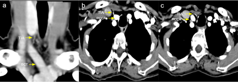
Objectives: The aim of this study is to determine the prevalence of thyroidea ima artery (TIA) and branching pattern variations of aortic arch among 2000 patients who underwent CECT imaging.
Materials and methods: This retrospective cross-sectional observational study included 2000 patients who underwent CECT of aortic arch and branches during a two years period in a tertiary care centre in South India. The presence of thyroidea ima artery with its origin were identified and the branching patterns of aortic arch were assessed.
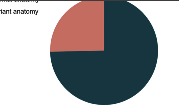
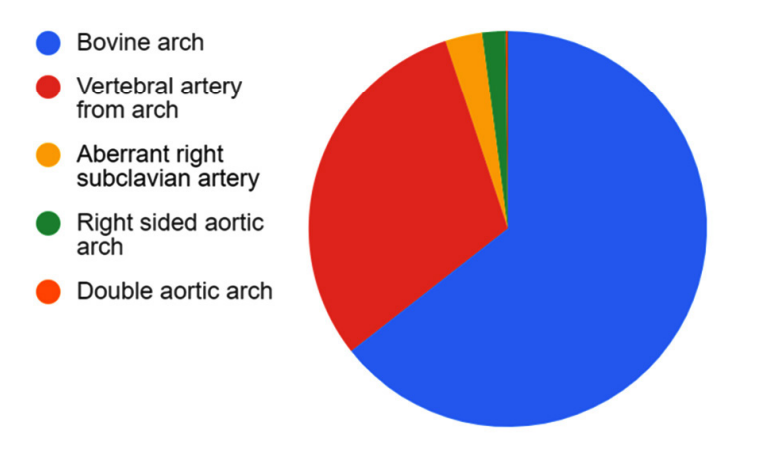
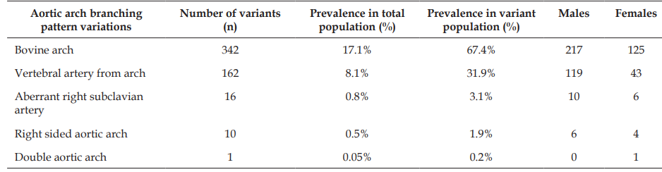
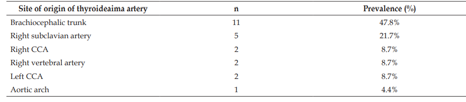
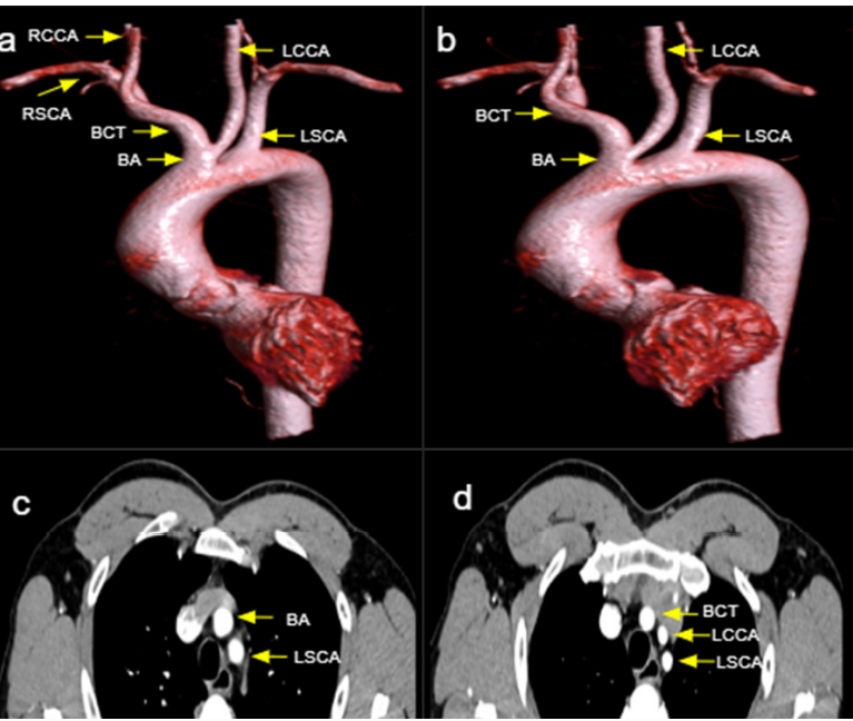
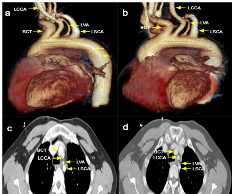
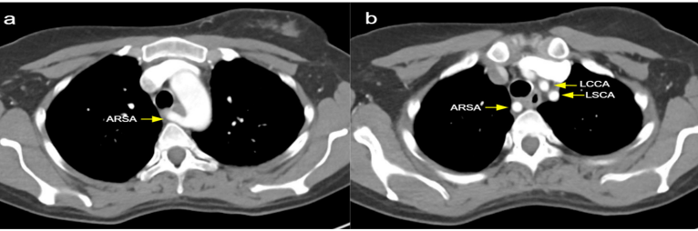
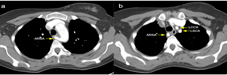
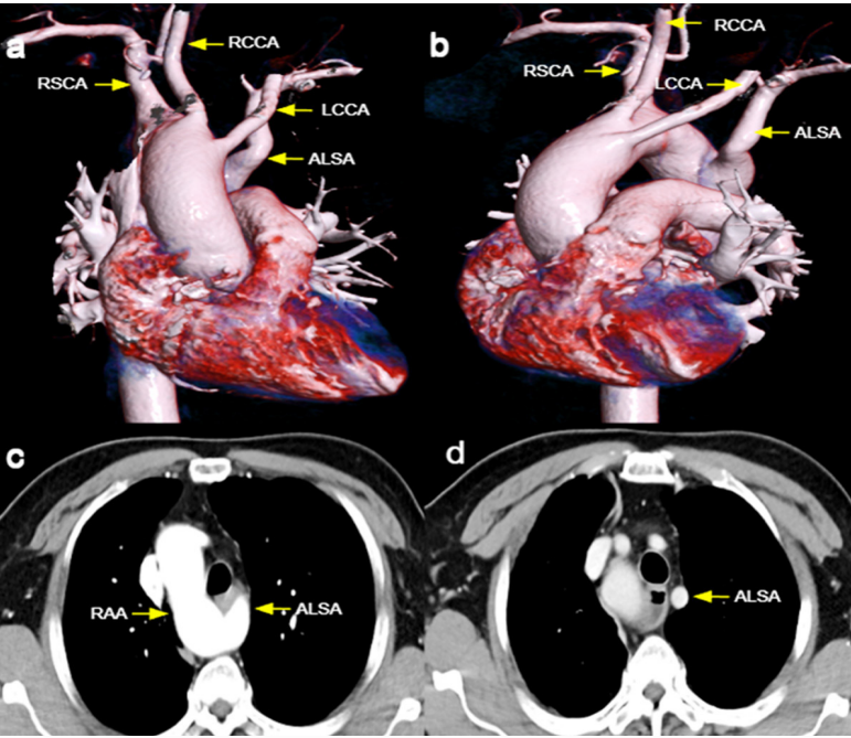
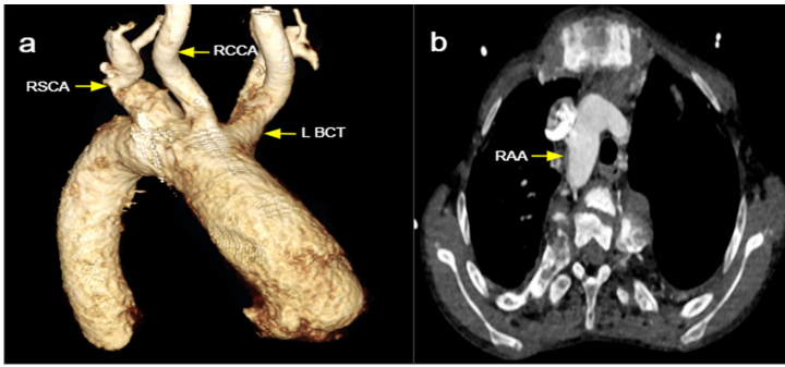
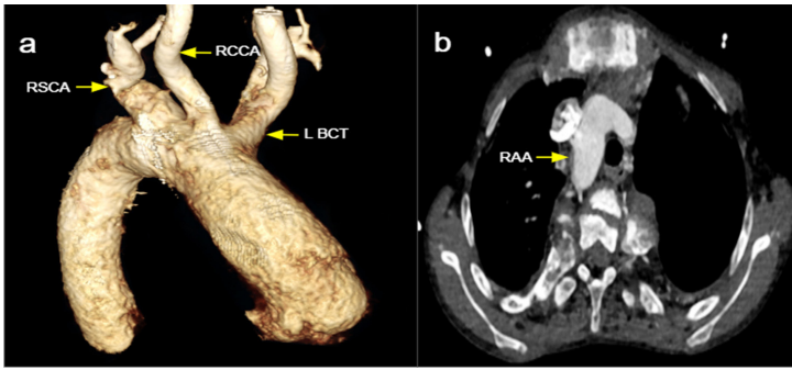
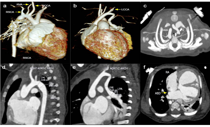
Results: TIA was identified in 23 patients, showing a prevalence of 1.15%. The normal branching pattern of aortic arch was observed in 1493 patients, and variations in branching pattern were seen in 507 patients. The variations included 342 cases of bovine type arch, 162 left vertebral arteries originating from arch, 16 aberrant right subclavian artery, 10 right sided aortic arch, and 1 double aortic arch.
Conclusion: The identification of TIA and variations in aortic arch branching are crucial, as they can cause complications during neck and thoracic surgeries and endovascular interventions. CECT is a reliable tool to assess the vascular anatomy. Considering the prevalence demonstrated in our study, imaging should be included prior to procedures in order to prevent complications.
References
- 1. Yurasakpong L, Nantasenamat C, Janta S, Eiamratchanee P, Coey J, Chaiyamoon A, et al. The decreasing prevalence of the thyroid ima artery: a systematic review and machine learning assisted meta-analysis. Ann Anat. 2022;239:151803.
- 2. Branca JJV, Lascialfari Bruschi A, Pilia AM, Carrino D, Guarnieri G, Gulisano M, et al. The thyroid gland: a revision study on its vascularization and surgical implications. Medicina (Kaunas). 2022;58(1):137.
- 3. Schleich JM. Development of the human heart: days 15–21. Heart. 2002;87(5):487–8.
- 4. Alsaif HA, Ramadan WS. An anatomical study of the aortic arch anomalies. J King Abdulaziz Univ Med Sci. 2010;17:37–54.
- 5. Jakanani GC, Adair W. Frequency of variations in aortic arch anatomy depicted on multidetector CT. Clin Radiol. 2010;65(6):481–7.
- 6. Müller M, Schmitz BL, Pauls S, Schick M, Röhrer S, Kapapa T, et al. Variations of the aortic arch–a study on the most common branching patterns. Acta Radiol. 2011;52(7):738–42.
- 7. Donnelly LF, Fleck RJ, Pacharn P, Ziegler MA, Fricke BL, Cotton RT. Aberrant subclavian arteries: cross-sectional imaging findings in infants and children referred for evaluation of extrinsic airway compression. AJR Am J Roentgenol. 2002;178(5):1269–74.
- 8. Ramos-Duran L, Nance JW Jr, Schoepf UJ, Henzler T, Apfaltrer P, Hlavacek AM. Developmental aortic arch anomalies in infants and children assessed with CT angiography. AJR Am J Roentgenol. 2012;198(5):W466–74.
- 9. Türkvatan A, Büyükbayraktar FG, Ölçer T, Cumhur T. Congenital anomalies of the aortic arch: evaluation with the use of multidetector computed tomography. Korean J Radiol. 2009;10(2):176–84.
- 10. Shuford WH, Sybers RG, Edwards FK. The three types of right aortic arch. Am J Roentgenol Radium Ther Nucl Med. 1970;109(1):67–74.
- 11. Esen K, Ozgur A, Balci Y, Tok S, Kara E. Variations in the origins of the thyroid arteries on CT angiography. Jpn J Radiol. 2018;36(2):96–102.
- 12. Chin J, Zhou Y, Wan PJ, Lomiguen CM. The prevalence of thyroid ima artery and its clinical significance. Int J Otorhinolaryngol Head Neck Surg. 2019;5(4):845–9.
- 13. Gwilym S, Cooney A. Acute fatal haemorrhage during percutaneous dilatational tracheostomy. Br J Anaesth. 2004;92(2):298.
- 14. Kamparoudi P, Paliouras D, Gogakos AS, Rallis T, Schizas NC, Lazopoulos A, et al. Percutaneous tracheostomy—beware of the thyroidea-ima artery. Ann Transl Med. 2016;4(22):449.
- 15. Shaw JA, Gravereaux EC, Eisenhauer AC. Carotid stenting in the bovine arch. Catheter Cardiovasc Interv. 2003;60(4):566–9.
- 16. Layton KF, Kallmes DF, Cloft HJ, Lindell EP, Cox VS. Bovine aortic arch variant in humans: clarification of a common misnomer. AJNR Am J Neuroradiol. 2006;27(7):1541–2.
- 17. Baadh AS, Rockman CB, Mitnick RJ, Lim RP. Bovine arch and carotid artery atherosclerosis: are they related? Clin Imaging. 2014;38(5):681–5.
- 18. Moore KL, Persaud TVN, Tochia MG. The Developing Human: Clinically Oriented Embryology. 7th ed. Philadelphia: Saunders; 2003. p. 259–63.
- 19. Graham JM, Feliciano DV, Mattox KL, Beall AC Jr. Innominate vascular injury. J Trauma. 1982;22(8):647–55.
- 20. Bergman RA, Afifi AK, Miyauchi R. Illustrated encyclopedia of human anatomic variation [Internet]. 2000
- 21. Nayak SR, Pai MM, Prabhu LV, D'Costa S, Shetty P. Anatomical organization of aortic arch variations in the India: embryological basis and review. J Vasc Bras. 2006;5(2):95–100.
- 22. Satti SR, Cerniglia CA, Koenigsberg RA. Cervical vertebral artery variations: an anatomic study. AJNR Am J Neuroradiol. 2007;28(5):976–80.
- 23. Sukumaran TT, Pillay M, Gopalakrishnan A. An anomalous right subclavian artery with a retrotracheal course: a case report. J Clin Diagn Res. 2015;9(11):AD01–2.
- 24. Fehmi Yazıcıoğlu H, Sevket O, Akın H, Aygün M, Özyurt ON, Karahasanoğlu A. Aberrant right subclavian artery in Down syndrome fetuses. Prenat Diagn. 2013;33(3):209–13.
- 25. Hori D, Tanaka M, Yamaguchi A, Adachi H. Type A aortic dissection, right-sided aortic arch, and thoracic aortic aneurysm. Asian Cardiovasc Thorac Ann. 2009;17(6):640–2.
- 26. McElhinney DB, Hoydu AK, Gaynor JW, Spray TL, Goldmuntz E, Weinberg PM. Patterns of right aortic arch and mirror-image branching of the brachiocephalic vessels without associated anomalies. Pediatr Cardiol. 2001;22(4):285–91.
- 27. Backer CL, Mongé MC, Popescu AR, Eltayeb OM, Rastatter JC, Rigsby CK. Vascular rings. Semin Pediatr Surg. 2016;25(3):165–75.
- 28. Guo Q, Kong Y, Zeng S, Zhou J, Wang X, Shang Q, et al. Fetal double aortic arch: prenatal sonographic and postnatal computed tomography angiography features, associated abnormalities and clinical outcomes. BMC Pregnancy Childbirth. 2020;20(1):738.
- 29. Nagre SW, Kulkarni DV. Double aortic arch surgery. Indian J Vasc Endovasc Surg. 2015;2(3):118–20.
- 30. Lale P, Toprak U, Yagız G, Kaya T, Uyanık SA. Variations in the branching pattern of the aortic arch detected with computerized tomography angiography. Adv Radiol. 2014;2014:969728.
Data Sharing Statement
There are no additional data available. All raw data and code are available upon request.
Funding
This research received no funding.
Author Contributions
Whether all authors contributed significantly to the work and approve its publication.
Ethics Declaration
This article does not involve any human or animal subjects, and therefore does not require ethics approval.
Acknowledgements
We would like to express our gratitude to the patients, their families, and all those who have contributed to this study.
Conflicts of Interest
The authors report no conflicts of interest in this work.
About this article

Cite this article
Periaswamy G, Christopher N, Arunachalam VK, et al. Prevalence of thyroidea ima artery & variations in aortic arch branches in South Asian population: a retrospective CT based study. Indian J Anat. 2023;12(1):9–19
Licence:
Attribution-Non-commercial 4.0 International (CC BY-NC 4.0)This license enables reusers to distribute, remix, adapt, and build upon the material in any medium or format for noncommercial purposes only, and only so long as attribution is given to the creator.
| Received | Accepted | Published |
|---|---|---|
| September 03, 2023 | October 10, 2023 | November 30, 2023 |
DOI: https://doi.org/10.21088/ija.2320.0022.12123.1
Keywords
Thyroideaima arteryRight aortic archDouble Aortic archBovine type arch.Right aortic archDouble Aortic archBovine type arch.Search for Similar Articles
Similar Articles
- Supernumerary Pulmonary Vein: A Rare Anatomical Variation Observed During Cadave...
- Elongated Styloid Process: Anatomical Variations and Their Clinical Relevance: A...
- Bridging the Gap Between Dissection and Surgery: The Role of Soft Embalming in M...
- A Cadaveric Study of Retro Hepatic Segment of Inferior Vena Cava and Ostia Venae...
- Estimation of Stature from Forearm Length in Medical Undergraduate Students of a...
Article Level Metrics
Last UpdatedTuesday 22 September 2026, 04:29:57 (IST)
5059
Accesses
27
655
00
Citations
NA
NA
NA
Download citation
Article Keywords
Keyword Highlighting
Highlight selected keywords in the article text.
Timeline
| Received | September 03, 2023 |
| Accepted | October 10, 2023 |
| Published | November 30, 2023 |
licence
Attribution-Non-commercial 4.0 International (CC BY-NC 4.0)
This license enables reusers to distribute, remix, adapt, and build upon the material in any medium or format for noncommercial purposes only, and only so long as attribution is given to the creator.