Full Text (PDF)
Indian Journal of Anatomy 15(2):p 89-91, May-August 2026. | DOI: https://doi.org/10.21088/ija.2320.0022.15226.6
Case Report
Supernumerary Pulmonary Vein: A Rare Anatomical Variation Observed During Cadaveric Dissection
Vishal Ramesh Jasuja, Abhigyan Singh, Nikhil Gupta, Mamta Rani, Monika Srivastava
Author Information
Licence:
Attribution-Non-commercial 4.0 International (CC BY-NC 4.0)This license enables reusers to distribute, remix, adapt, and build upon the material in any medium or format for noncommercial purposes only, and only so long as attribution is given to the creator.
Indian Journal of Anatomy 15(2):p 89-91, May-August 2026. | DOI: https://doi.org/10.21088/ija.2320.0022.15226.6
How Cite This Article:
Abhigyan Singh, Nikhil Gupta, Mamta Rani et. al, Supernumerary Pulmonary Vein: A Rare Anatomical Variation Observed During Cadaveric Dissection. Ind Jr Anat. 2026; 15(2): 89-91.Timeline
Received : June 19, 2026
Accepted : July 18, 2026
Published : August 30, 2026
Abstract
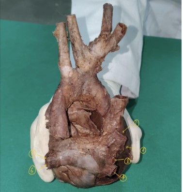
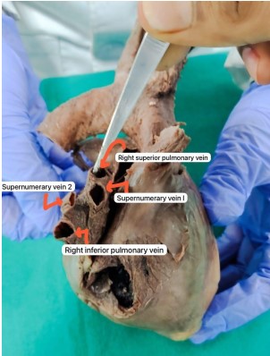
Introduction: Pulmonary veins are responsible for carrying oxygenated blood from the lungs to the left atrium of the heart. Unlike other veins, they supply oxygen rich blood to the left atrium of the heart. They have thin walls and are valveless. Normally, four pulmonary veins are present: Right Superior, Left Superior, Right Inferior, Left Inferior hence presence of a supernumerary pulmonary vein is considered a rare anatomical variation. Case Report: After careful removal of heart from thoracic cavity, We observed the presence of two additional veins alongside of the four above mentioned pulmonary veins. Upon further investigation we noted that they emerged from the right lung draining independently into the left atrium. Supernumerary veins were identified along with the normal superior and inferior pulmonary veins. Conclusion: This study strongly emphasizes the importance of keeping this anomaly in mind so that recognition by radiologists and surgeons can help prevent surgical complications and improve success of interventional procedures.
References
- 1. Standring S, ed. Gray's Anatomy: The Anatomical Basis of Clinical Practice. 41st ed. Philadelphia: Elsevier; 2016.
- 2. Moore KL, Persaud TVN, Torchia MG. The Developing Human: Clinically Oriented Embryology. 10th ed. Philadelphia: Elsevier; 2016.
- 3. Marom EM, Herndon JE, Kim YH, McAdams HP. Variations in pulmonary venous drainage to the left atrium: implications for radiofrequency ablation. Radiology. 2004;230(3):824-829.
- 4. Cronin P, Sneider MB, Kazerooni EA, et al. MDCT evaluation of pulmonary venous anatomy. AJR Am J Roentgenol. 2007;188(4):W419-W425.
- 5. Sobrinho G, Almeida J, et al. Anatomical variations of pulmonary veins: imaging findings and clinical implications. Surg Radiol Anat. 2013;35(7):567-574.
- 6. Calkins H, Brugada J, Packer DL, et al. HRS/ EHRA/ECAS expert consensus statement on catheter and surgical ablation of atrial fibrillation. Heart Rhythm. 2007;4(6):816-861.
- 7. Webb WR, Higgins CB. Thoracic Imaging: Pulmonary and Cardiovascular Radiology. 2nd ed. Philadelphia: Lippincott Williams & Wilkins; 2010.
- 8. Hollinshead WH. Anatomy for Surgeons. Vol 2. New York: Harper & Row; 1968.
- 9. Snell RS. Clinical Anatomy by Regions. 10th ed. Philadelphia: Wolters Kluwer; 2019.
- 10. Ho SY, Cabrera JA, Sanchez-Quintana D. Architecture of the pulmonary veins: relevance to radiofrequency ablation. Heart. 2001;86(3):265-270.
Data Sharing Statement
There are no additional data available. All raw data and code are available upon request.
Funding
This research received no funding.
Author Contributions
All authors contributed significantly to the work and approve its publication.
Ethics Declaration
This article does not involve any human or animal subjects, and therefore does not require ethics approval.
Acknowledgements
We would like to express our gratitude to the patients, their families, and all those who have contributed to this study.
Conflicts of Interest
No conflicts of interest in this work.
About this article

Cite this article
Abhigyan Singh, Nikhil Gupta, Mamta Rani et. al, Supernumerary Pulmonary Vein: A Rare Anatomical Variation Observed During Cadaveric Dissection. Ind Jr Anat. 2026; 15(2): 89-91.
Licence:
Attribution-Non-commercial 4.0 International (CC BY-NC 4.0)This license enables reusers to distribute, remix, adapt, and build upon the material in any medium or format for noncommercial purposes only, and only so long as attribution is given to the creator.
| Received | Accepted | Published |
|---|---|---|
| June 19, 2026 | July 18, 2026 | August 30, 2026 |
DOI: https://doi.org/10.21088/ija.2320.0022.15226.6
Keywords
Pulmonary VeinSupernumerary Pulmonary VeinLeft AtriumAnatomical VariationCadaveric Study.Search for Similar Articles
Similar Articles
- Elongated Styloid Process: Anatomical Variations and Their Clinical Relevance: A...
- Bridging the Gap Between Dissection and Surgery: The Role of Soft Embalming in M...
- A Cadaveric Study of Retro Hepatic Segment of Inferior Vena Cava and Ostia Venae...
- Estimation of Stature from Forearm Length in Medical Undergraduate Students of a...
- Morphological Variations of Gall-bladder: A Cadaveric and Ultrasonographic Study
Article Level Metrics
Last UpdatedTuesday 29 September 2026, 12:06:54 (IST)
5321
Accesses
66
664
00
Citations
NA
NA
NA
Download citation
Article Keywords
Keyword Highlighting
Highlight selected keywords in the article text.
Timeline
| Received | June 19, 2026 |
| Accepted | July 18, 2026 |
| Published | August 30, 2026 |
licence
Attribution-Non-commercial 4.0 International (CC BY-NC 4.0)
This license enables reusers to distribute, remix, adapt, and build upon the material in any medium or format for noncommercial purposes only, and only so long as attribution is given to the creator.