Full Text (PDF)
Indian Journal of Anatomy 15(2):p 47-52, May-August 2026. | DOI: https://doi.org/10.21088/ija.2320.0022.15226.1
Original Article
Morphological Variations of Gall-bladder: A Cadaveric and Ultrasonographic Study
Sharada B Menasinkai, Yashas N Hegde
Author Information
Licence:
Attribution-Non-commercial 4.0 International (CC BY-NC 4.0)This license
enables reusers to distribute, remix, adapt, and build upon the material in any
medium or format for noncommercial purposes only, and only so long as
attribution is given to the creator.
Indian Journal of Anatomy 15(2):p 47-52, May-August 2026. | DOI: https://doi.org/10.21088/ija.2320.0022.15226.1
How Cite This Article:
Yashas N Hegde, Sharada B Menasinkai. Morphological Variations of Gall-bladder: A Cadaveric and Ultrasonographic Study. Ind Jr Anat. 2026; 15(2): 47-52.Timeline
Received : March 31, 2026
Accepted : June 20, 2026
Published : August 30, 2026
Abstract
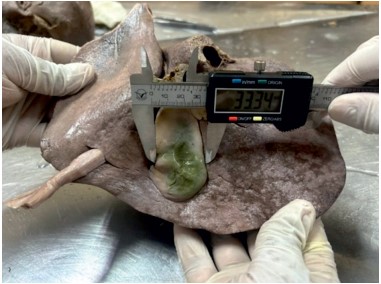
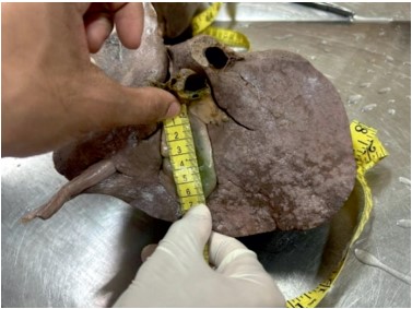
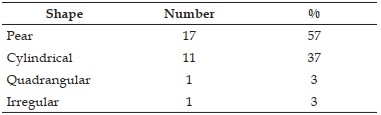
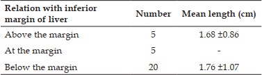
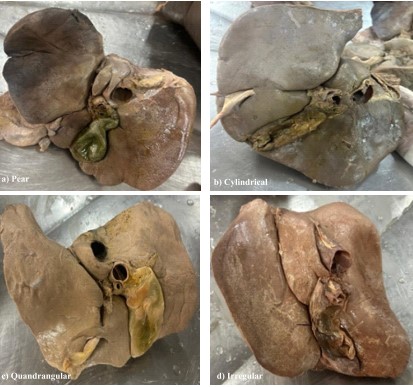
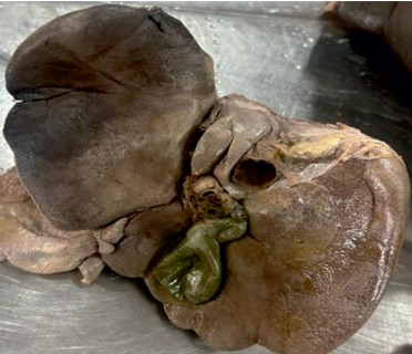
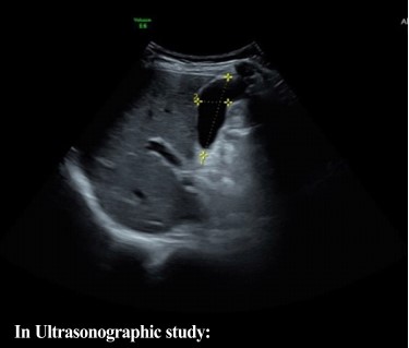
Introduction: The gall bladder is an important component of the extrahepatic biliary apparatus which presents various morphological variations, the knowledge of which serves to be important during radiological scans and surgeries. The study aims to identify few anatomical variations in the dimensions, shape and position of gall bladder which would serve beneficial in the above mentioned procedures as well as in other anatomical, clinical and research settings. Methodology: The study comprised of 30 thoroughly dissected Liver specimens obtained from 10% formalin fixed cadavers of both genders and 20 ultrasonographic scans of abdomen from the Department of Anatomy and Department of Radiology, AIMS after the requested consent from Institutional Ethics Committee and the Head of Department. The study carried out for a period of four months from February 2025 to June 2025 considered the parameters length, breadth, shape of gall bladders and length of part below or above inferior border of liver for cadaveric specimens. Results: The mean length, breadth of gall bladders in cadaveric and ultrasonographic studies were 7.82±1.29, 3.66±0.81, 6.155±0.84 and 2.39±0.59. 20 gall bladders were below the inferior margin of liver (mean length = 1.76±1.07), 5 gall bladders were above the margin (mean length = 1.68±0.86) and 5 gall bladders were at the margin. The shapes observed were pear (57%), cylindrical (37%), quadrangular (3%) and others (3%). Conclusion: The study would serve important for working surgeons and radiologists treating and scanning cases of gall bladder diseases.
References
- 1. Standring S. Gray’s anatomy: The anatomical basis of the clinical practice, 40 Edition. Elsevier Churchill Livingstone, Philadelphia, London 2008;1177-1181.
- 2. Singh VK, Hussain MZ, Kumar R, Prasad R. A Morphometric Study of the Adult Human Gall Bladder in a Cadaver and its Clinical Significance in the Eastern Indian Population. Int J Pharm Clin Res. 2022;14(4):605-10.
- 3. Nayak G, Sahoo N, Panda SK. Study of Morphological Variations of Gallbladder in Eastern Indian Cadavers. J Krishna Inst Med Sci Univ 2021; 10(4):56-63.
- 4. Srivastava S, Khan AZ. Morphology of Gallbladder – A Cadaveric Study. J Curr Med Res Opin. 2019;2(10):293–8.
- 5. Divya C, Ashwini NS, Sridevi NS. Morphometric study of gallbladder in cadavers. J Res Appl Basic Med Sci. 2022;8(1):8–14.
- 6. D D Neginhal, U K Kulkarni. Morphology of gallbladder - A cadaveric study. MedPulse – International Journal of Anatomy. December 2018; 8(3): 44-47.
- 7. Siva Prasad Chavva, Santosh U Kanpur. A study of sonographic assessment of gallbladder dimensions in normal adults. International Journal of Contemporary Medicine Surgery and Radiology. 2018;3(4): D157-D159.
- 8. Adeyekun AA, Ukadike IO. Sonographic evaluation of gallbladder dimensions in healthy adults in Benin City, Nigeria. West Afr J Radiol. 2013;20(1):4–8.
- 9. Jamal AF, Muhammad BS, Rashid SA. Ultrasound evaluation of gallbladder dimensions in healthy adults. Zanco J Med Sci. 2022;26(1):88–95.
- 10. Rumack CM, Levine D. The biliary tree and gallbladder. In: Rumack CM, Levine D, editors. Diagnostic ultrasound. 6th ed. Vol. 1. Philadelphia: Elsevier; 2024. p. 228–229.
Data Sharing Statement
There are no additional data available. All raw data and code are available upon request.
Funding
This research received no funding.
Author Contributions
All authors contributed significantly to the work and approve its publication.
Ethics Declaration
This article does not involve any human or animal subjects, and therefore does not require ethics approval.
Acknowledgements
We would like to express our gratitude to the patients, their families, and all those who have contributed to this study.
Conflicts of Interest
No conflicts of interest in this work.
About this article

Cite this article
Yashas N Hegde, Sharada B Menasinkai. Morphological Variations of Gall-bladder: A Cadaveric and Ultrasonographic Study. Ind Jr Anat. 2026; 15(2): 47-52.
Licence:
Attribution-Non-commercial 4.0 International (CC BY-NC 4.0)This license
enables reusers to distribute, remix, adapt, and build upon the material in any
medium or format for noncommercial purposes only, and only so long as
attribution is given to the creator.
| Received | Accepted | Published |
|---|---|---|
| March 31, 2026 | June 20, 2026 | August 30, 2026 |
DOI: https://doi.org/10.21088/ija.2320.0022.15226.1
Keywords
Gall BladderUltrasonographyMorphologySearch for Similar Articles
Similar Articles
- Supernumerary Pulmonary Vein: A Rare Anatomical Variation Observed During Cadave...
- Elongated Styloid Process: Anatomical Variations and Their Clinical Relevance: A...
- Bridging the Gap Between Dissection and Surgery: The Role of Soft Embalming in M...
- A Cadaveric Study of Retro Hepatic Segment of Inferior Vena Cava and Ostia Venae...
- Estimation of Stature from Forearm Length in Medical Undergraduate Students of a...
Article Level Metrics
Last UpdatedMonday 07 September 2026, 23:19:24 (IST)
4699
Accesses
30
516
00
Citations
NA
NA
NA
Download citation
Article Keywords
Keyword Highlighting
Highlight selected keywords in the article text.
Timeline
| Received | March 31, 2026 |
| Accepted | June 20, 2026 |
| Published | August 30, 2026 |
licence
Attribution-Non-commercial 4.0 International (CC BY-NC 4.0)
This license
enables reusers to distribute, remix, adapt, and build upon the material in any
medium or format for noncommercial purposes only, and only so long as
attribution is given to the creator.