Full Text (PDF)
Indian Journal of Pathology: Research and Practice 13(3):p 107-110, July- September 2024. | DOI: https://doi.org/10.21088/ijprp.2278.148X.13324.3
Case Report
Placental Chorioangioma Cellular Histological Subtype: A Rare Primary Benign Tumor of Placenta
Sunil V. Jagtap, Shashwita S. Jagtap, Neha N. Ghadge, Manasi R. Bhade, Pooja Pandey, Abhilasha S. Jadhav
Author Information
Licence:
Attribution-Non-commercial 4.0 International (CC BY-NC 4.0)This license enables reusers to distribute, remix, adapt, and build upon the material in any medium or format for noncommercial purposes only, and only so long as attribution is given to the creator.
Indian Journal of Pathology: Research and Practice 13(3):p 107-110, July- September 2024. | DOI: https://doi.org/10.21088/ijprp.2278.148X.13324.3
How Cite This Article:
Sunil V. Jagtap, Shashwita S. Jagtap, Neha N. Ghadge et al. Placental Chorioangioma Cellular Histological Subtype: A Rare Primary Benign Tumor of Placenta. Ind Jr of Path: Res and Practice 2024;13(3)107-110.Timeline
Received : July 12, 2024
Accepted : August 31, 2024
Published : September 30, 2024
Abstract
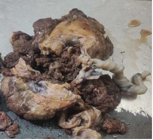
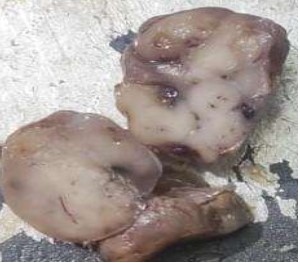
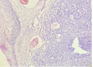
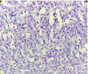
Background: Placental chorangioma is a benign angioma arising from chorionic tissue. These are well demarcated placental mass composed of capillaries, stromal cells and surrounding trophoblast arising in a stem villus. Case report: A 28-year-old, gravida 2 para 1, L1 was admitted for abdominal pain, vaginal bleeding and foul smelling discharge. She had history of normal vaginal delivery at home two month back. On per vaginal examination cervix dilated, foul smelling discharge was noted. Clinically suspected of retained placenta, placenta increta. USG abdomen and pelvis showed an enlarged uterus with a focal heterogeneously hyperechoic lesion measures 44x23x34mm noted in the upper endometrial cavity. Moderate increase vascularity in color doppler examination. The findings likely placental remains (? Placenta increta) / retained product of conceptions. No pelvic mass was seen. Hysterecscopy with dilation and curettage were done for removal of retained products. On gross pathological examination single, rounded, well demarcated placental mass was seen. It was firm, reddish brown in color with localization on the fetal side of the placental disk. On histopathology reported as placental chorangioma cellular subtype. Chorioamnitis was noted. Areas of degenerative changes, necrosis, focal calcification, and hyalinization was noted. Conclusion: Placental chorioangioma is a rare benign tumor of the placenta. Early diagnosis, close prenatal checkup and monitor with appropriate intervention may prevent severe fetal and maternal complications and perinatal mortality related to chorioangioma.
References
- 1. Fox H. Non-trophoblastic tumour of the placenta. In: Fox H, Wells M, editors. Haines and Taylor Obstetrical and Gynecological Pathology. 5th ed. Edinburgh: Churchill Livingstone; 2003. p. 1449-63.
- 2. Guschmann, M., Henrich, W., Entezami, M. and Dudenhausen, J. W. Chorioangioma – new insights into a well-known problem I. Results of a clinical and morphological study of 136 cases. 2003,31, 2: 163-169.
- 3. Wou K, Chen MF, Mallozzi A, Brown RN, Shrim A Pregnancy outcomes and ultrasonographic diagnosis in patients with histologically-proven placental chorioangioma Placenta 2011, 32. 671- 4.
- 4. Singh A, Sivaranjani, Bagga R, Saha PK, Dey P. Successful outcome of giant chorioangioma. J Family Med Prim Care. 2021 May;10(5):2038-2040.
- 5. Lež C, Fures R, Hrgovic Z, Belina S, Fajdic J, Münstedt K. “Chorangioma placentae”. Rare Tumors.2010, 2 (4): e67.
- 6. Bashiri A, Furman B, Erez O, Wiznitzer A, Holcberg G, Mazor M. Twelve cases of placental chorioangioma. Pregnancy outcome and clinical significance. Arch Gynecol Obstet. 2002;266(1):53-5.
- 7. Jauniaux, E, Ogle, R. Color Doppler imaging in the diagnosis and management of chorioangiomas. Ultrasound Obstet Gynecol 2000;15:463–7.
- 8. Marchetti AA. A consideration of certain types of benign tumors of the placenta. Surgery, Gynecology and Obstetrics. 1939;68:733–743.
- 9. Rech F, Salernitano D, Patella A. Placental chorioangioma. Minerva Ginecol 2002;54:417-33.
- 10. Jagtap SV, Aher V, Gadhiya S, Jagtap SS. Gestational Trophoblastic Disease - Clinicopathological Study at Tertiary Care Hospital. J Clin Diagn Res. 2017 Aug;11(8):EC27-EC30.
- 11. Ogino S, Redline RW. Villous capillary lesions of the placenta: Distinctions between chorangioma, chorangiomatosis, and chorangiosis. Hum Pathol 2000;31:945-54.
Data Sharing Statement
There are no additional data available. All raw data and code are available upon request.
Funding
This research received no funding.
Author Contributions
All authors contributed significantly to the work and approve its publication.
Ethics Declaration
This article does not involve any human or animal subjects, and therefore does not require ethics approval.
Acknowledgements
We would like to express our gratitude to the patients, their families, and all those who have contributed to this study.
Conflicts of Interest
No conflicts of interest in this work.
About this article

Cite this article
Sunil V. Jagtap, Shashwita S. Jagtap, Neha N. Ghadge et al. Placental Chorioangioma Cellular Histological Subtype: A Rare Primary Benign Tumor of Placenta. Ind Jr of Path: Res and Practice 2024;13(3)107-110.
Licence:
Attribution-Non-commercial 4.0 International (CC BY-NC 4.0)This license enables reusers to distribute, remix, adapt, and build upon the material in any medium or format for noncommercial purposes only, and only so long as attribution is given to the creator.
| Received | Accepted | Published |
|---|---|---|
| July 12, 2024 | August 31, 2024 | September 30, 2024 |
DOI: https://doi.org/10.21088/ijprp.2278.148X.13324.3
Keywords
HemangiomaTumors of placentaNon-trophoblastic tumorsPlacental Chorioangioma PlacentaSearch for Similar Articles
Similar Articles
- Clinico-Pathological and Immunohistochemical Study of Primary CNS Tumors in a Te...
- The Role of SGLT2 Inhibitors in Cardiovascular Protection Beyond Diabetes: A Co...
- Epidermal Inclusion Cyst Presenting as 4th Ventricle Space Occupying Lesion: A R...
- Silent Retroperitoneal Seminoma Arising from an Occult Undescended Testis: Diagn...
- Scapular Metastasis of Oral Squamous Cell Carcinoma: A Case Report of an Unusual...
Article Level Metrics
Last UpdatedTuesday 22 September 2026, 04:35:10 (IST)
3671
Accesses
12
541
00
Citations
NA
NA
NA
Download citation
Article Keywords
Keyword Highlighting
Highlight selected keywords in the article text.
Timeline
| Received | July 12, 2024 |
| Accepted | August 31, 2024 |
| Published | September 30, 2024 |
licence
Attribution-Non-commercial 4.0 International (CC BY-NC 4.0)
This license enables reusers to distribute, remix, adapt, and build upon the material in any medium or format for noncommercial purposes only, and only so long as attribution is given to the creator.