Full Text (PDF)
Indian Journal of Pathology: Research and Practice 13(3):p 111-114, July- September 2024. | DOI: https://doi.org/10.21088/ijprp.2278.148X.13324.4
Case Report
Papillary Tumor of Pineal Region: Clinical Manifestations, Histopathological Insights, and Immunohistochemical Profile: A Comprehensive Case Report
Singh A, Bharti S, Guralarasan G, Bhadani PP, Kumari M
Author Information
Licence:
Attribution-Non-commercial 4.0 International (CC BY-NC 4.0)This license
enables reusers to distribute, remix, adapt, and build upon the material in any
medium or format for noncommercial purposes only, and only so long as
attribution is given to the creator.
Indian Journal of Pathology: Research and Practice 13(3):p 111-114, July- September 2024. | DOI: https://doi.org/10.21088/ijprp.2278.148X.13324.4
How Cite This Article:
Alankrita Madhur, Alpana Jain, Alka Mittal. Hydatid Cyst in Breast: A Rare Case Report with Review Literature. Ind Jr of Path: Res and Practice 2024;13(3)111-114.Timeline
Received : August 03, 2024
Accepted : September 05, 2024
Published : September 30, 2024
Abstract
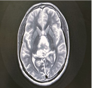
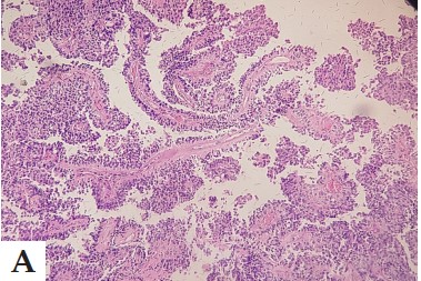
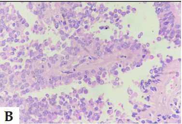
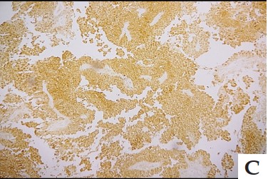
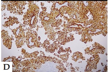
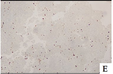
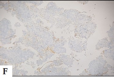
Introduction: Papillary tumor of the pineal region (PTPR) is a unique rare neuroepithelial tumor arising from unique ependymocytes of the subcommissural organ, different from the pineal gland Affecting less than 1% of adult intracranial tumors, PTPR presented unique diagnostic challenges due to rarely and because of its nature papillary pattern does. Case presentation: We report a 25-year-old man initially treated for acute hydrocephalus and later diagnosed with PTPR. Radiologic examination revealed a typical pineal area mass, and histopathology examination confirmed PTPR with prominent papillary structures. Immunohistochemistry played an important role in supporting the disease. Conclusion: This article outlines a complex approach to the diagnosis of PTPR, including radiology, histopathology, and immunohistochemistry. Furthermore, it highlights the uniqueness of PTPR in pineal-parenchymal tumor classification, as outlined in the WHO classification for 2021. This report contributes to the paucity of literature on this rare tumor, and highlights comprehensive diagnostic approach when evaluating pineal region masses.
References
- 1. Jouvet A, Fauchon F, Liberski P, Saint-Pierre G, Didier-Bazes M, Heitzmann A, et al. Papillary tumor of the pineal region. Am J Surg Pathol. 2003 Apr;27(4):505–12.
- 2. Louis DN, Perry A, Wesseling P, Brat DJ, Cree IA, Figarella-Branger D, et al. The 2021 WHO Classification of Tumors of the Central Nervous System: a summary. Neuro-Oncol. 2021 Aug 1;23(8):1231–51.
- 3. Roncaroli F, Scheithauer BW. Papillary Tumor of the Pineal Region and Spindle Cell Oncocytoma of the Pituitary: New Tumor Entities in the 2007 WHO Classification. Brain Pathol. 2007 Jun 26;17(3):314–8.
- 4. Hasselblatt M, Blümcke I, Jeibmann A, Rickert CH, Jouvet A, van de Nes J a. P, et al. Immunohistochemical profile and chromosomal imbalances in papillary tumours of the pineal region. Neuropathol Appl Neurobiol. 2006 Jun;32(3):278–83.
- 5. Kawahara I, Tokunaga Y, Yagi N, Iseki M, Abe K, Hayashi T. Papillary tumor of the pineal region. Neurol Med Chir (Tokyo). 2007 Dec;47(12):568–71.
- 6. Shibahara J, Todo T, Morita A, Mori H, Aoki S, Fukayama M. Papillary neuroepithelial tumor of the pineal region. A case report. Acta Neuropathol (Berl). 2004 Oct;108(4):337–40.
- 7. Fèvre Montange M, Vasiljevic A, Bergemer Fouquet AM, Bernier M, Champier J, Chrétien F, et al. Histopathologic and ultrastructural features and claudin expression in papillary tumors of the pineal region: a multicenter analysis. Am J Surg Pathol. 2012 Jun;36(6):916–28.
- 8. Fèvre-Montange M, Hasselblatt M, FigarellaBranger D, Chauveinc L, Champier J, Saint-Pierre G, et al. Prognosis and histopathologic features in papillary tumors of the pineal region: a retrospective multicenter study of 31 cases. J Neuropathol Exp Neurol. 2006 Oct;65(10)
Data Sharing Statement
There are no additional data available. All raw data and code are available upon request.
Funding
This research received no funding.
Author Contributions
All authors contributed significantly to the work and approve its publication.
Ethics Declaration
This article does not involve any human or animal subjects, and therefore does not require ethics approval.
Acknowledgements
We would like to express our gratitude to the patients, their families, and all those who have contributed to this study.
Conflicts of Interest
No conflicts of interest in this work.
About this article

Cite this article
Alankrita Madhur, Alpana Jain, Alka Mittal. Hydatid Cyst in Breast: A Rare Case Report with Review Literature. Ind Jr of Path: Res and Practice 2024;13(3)111-114.
Licence:
Attribution-Non-commercial 4.0 International (CC BY-NC 4.0)This license
enables reusers to distribute, remix, adapt, and build upon the material in any
medium or format for noncommercial purposes only, and only so long as
attribution is given to the creator.
| Received | Accepted | Published |
|---|---|---|
| August 03, 2024 | September 05, 2024 | September 30, 2024 |
DOI: https://doi.org/10.21088/ijprp.2278.148X.13324.4
Keywords
Brain neoplasmsPapillary tumors of the pineal regionPineal parenchymal tumorSearch for Similar Articles
Similar Articles
- Clinico-Pathological and Immunohistochemical Study of Primary CNS Tumors in a Te...
- The Role of SGLT2 Inhibitors in Cardiovascular Protection Beyond Diabetes: A Co...
- Epidermal Inclusion Cyst Presenting as 4th Ventricle Space Occupying Lesion: A R...
- Silent Retroperitoneal Seminoma Arising from an Occult Undescended Testis: Diagn...
- Scapular Metastasis of Oral Squamous Cell Carcinoma: A Case Report of an Unusual...
Article Level Metrics
Last UpdatedTuesday 22 September 2026, 01:54:08 (IST)
3670
Accesses
7
541
00
Citations
NA
NA
NA
Download citation
Article Keywords
Keyword Highlighting
Highlight selected keywords in the article text.
Timeline
| Received | August 03, 2024 |
| Accepted | September 05, 2024 |
| Published | September 30, 2024 |
licence
Attribution-Non-commercial 4.0 International (CC BY-NC 4.0)
This license
enables reusers to distribute, remix, adapt, and build upon the material in any
medium or format for noncommercial purposes only, and only so long as
attribution is given to the creator.