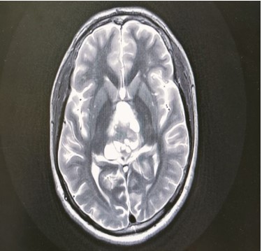
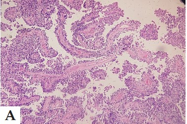
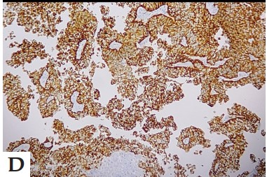
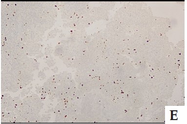
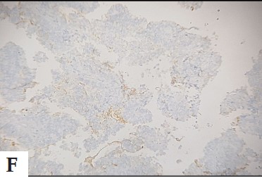
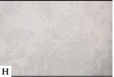
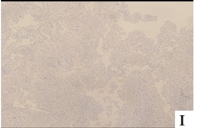
Description: Photomicrograph showing MRI brain contrast with MR spectroscopy, a large, measuring 4.4X3.0X4.6 cm (APXTRXCC) well-circumscribed mass was seen epicentred in the pineal gland and third ventricle. On post-contrast, avid enhancement of the solid component is seen.
Heading
Description: No description available.
Heading
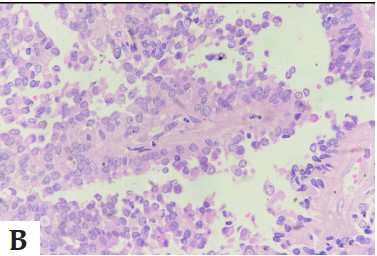
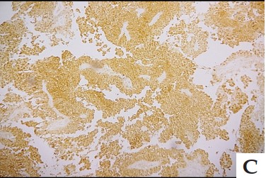
Description: These tumour cells are cuboidal to columnar, have mild pleomorphism, round to ovoid nuclei, fine chromatin, and moderately eosinophilic to occasional vacuolated cytoplasm (H&E 400x). On IHC, the tumour cells are positive for