Full Text (PDF)
Indian Journal of Forensic Medicine and Pathology 13(1):p 9-13, January-March 2020. | DOI: https://doi.org/10.21088/ijfmp.0974.3383.13120.1
Original Article
Patterns of Lung Lesions in Autopsy: A Histopathological Study
Anisha TS, Shashikala K2, Ramya T null
Author Information
Licence:
Attribution-Non-commercial 4.0 International (CC BY-NC 4.0)This license enables reusers to distribute, remix, adapt, and build upon the material in any medium or format for noncommercial purposes only, and only so long as attribution is given to the creator
Indian Journal of Forensic Medicine and Pathology 13(1):p 9-13, January-March 2020. | DOI: https://doi.org/10.21088/ijfmp.0974.3383.13120.1
How Cite This Article:
AnishaTS, Shashikala K, Ramya T et al. Patterns of Lung Lesions in Autopsy: A Histopathological Study. Indian J Forensic Med Pathol. 2020;13(1):9-13
Timeline
Received : January 02, 2020
Accepted : February 02, 2020
Published : March 30, 2020
Abstract
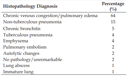
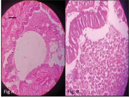
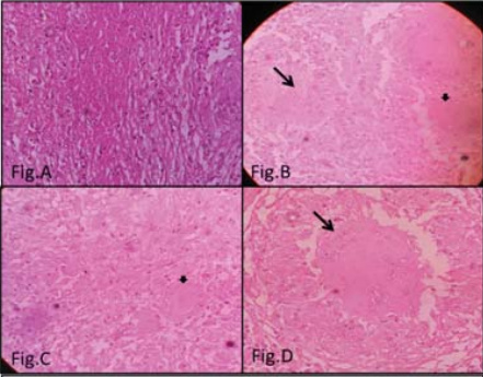
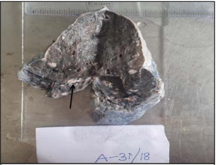
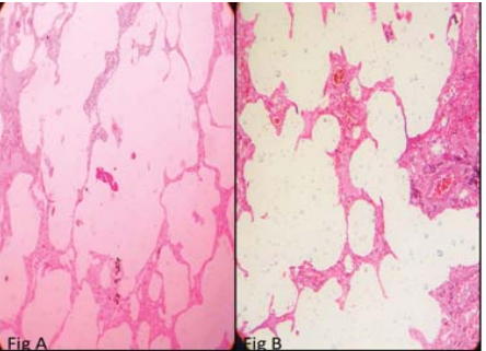
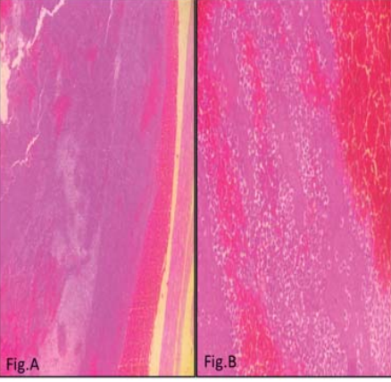
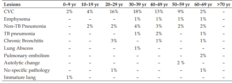
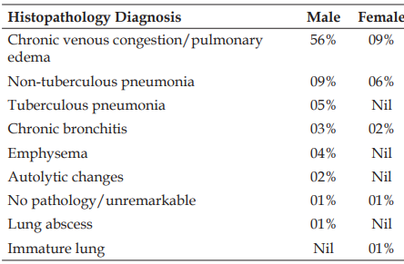
Introdution: Autopsy is recognized as a necessary part of medicine to establish final diagnosis, and relate the cause of death to the associated pathologies and the interaction between the two. Aim: To study and highlight the histopathological changes in lungs seen in autopsy cases. Methods: A cross-sectional study was done in the department of Pathology in a tertiary care hospital in Bangalore. A total of 100 lung specimens received from the department of forensic medicine for clinical autopsy over 3 years (January 2016 to January 2019) were studied. After noting the patient details and autopsy findings, the specimens were examined grossly and based on the gross findings, representative bits were given for histopathological examination. The histopathological findings were noted and results were statistically analyzed. Results: Amongst the 100 lungs studied, the commonest lesion was Congestion and/ Pulmonary edema (64%), followed by non-tuberculous pneumonia (15%), chronic bronchitis (5%), emphysema (4%), tuberculous pneumonia (4%), autolytic changes (2%), pulmonary embolism (2%), lung abscess (1%) and an immature lung (1%). Conclusion: This study highlights the various lesions in lungs from patients with history of varied causes of death and the lung lesions contributing directly or indirectly to the cause of death.
References
- 1. Costache M, Lazaroiu AM, Contolenco A, et al. Clinical or postmortem? The importance of the autopsy: a retrospective study. Maedica (Bucur). 2014;9(3):261–5.
- 2. Kotabagi RB, Charati SC, Jayachandar D. Clinical autopsy vs medicolegal autopsy. Med J Armed Forces India. 2005;61(3):258–63.
- 3. Kumar V, Abbas AK, Aster JC. Robbins and Cotran pathologic basis of disease. South Asia ed. Vol 2. New Delhi: Elsevier; 2014.
- 4. Patel CB, Patel K, Bhagat VM, et al. Pattern of histopathological lesions in lung autopsy. Int J Res Med Sci. 2018;6(1):279–83.
- 5. Amin NS, Shah PY, Patel RG, et al. Histopathological alterations in lung tissue received as autopsy specimens–a study of 410 cases. Int J Med Sci Public Health. 2017;6(1):327–30.
- 6. Chakma P, Thounaojam MD, Devi M. Incidental lung pathologies in medicolegal autopsies: a study. Indian J Forensic Community Med. 2017;4(3):189–94.
- 7. Hanmante RD, Chavan YH, Mulay PS, et al. Histopathological patterns of lung lesions in autopsy cases. Int J Adv Health Sci. 2014;1(1):15–9.
- 8. Selvam VR, Selvi T, Subramaniam PM. Prevalence of common disease in lungs and liver: a histopathological study. J Pharm Biomed Sci. 2011;12(9):1–5.
- 9. Mangal K, Yadav M. Incidence of liver diseases: a retrospective study of 1348 autopsy cases at tertiary care centre Jaipur. Int J Curr Res. 2015;7(12):23725–9.
- 10. Chauhan G, Agrawal M, Thakkar N, et al. Spectrum of histopathological lesions in lung autopsy. J Res Med Dent Sci. 2015;3(2):109–12.
- 11. Kurawar RR, Vasaikar MS. Spectrum of histomorphological changes in lungs at autopsy: a 5-year study. Ann Pathol Lab Med. 2017;4(1):A106–12.
- 12. Kurawar RR, Vasaikar MS. Spectrum of histomorphological changes in lungs at autopsy: a 5-year study. Ann Pathol Lab Med. 2017;4(1):A106–12.(Note: Item 12 in your source list appears to be a duplicate of Item 11; I have formatted both identically according to the correct journal standards.)
- 13. Bal MS, Sethi PS, Suri AK, et al. Histopathological pattern in lung autopsies. J Punjab Acad Forensic Med Toxicol. 2008;8(2):29–31.
- 14. Khare P, Gupta R, Ahuja M, et al. Prevalence of lung lesions at autopsy: a histopathological study. J Clin Diagn Res. 2017;11(5):EC13–6.
Data Sharing Statement
There are no additional data available. All raw data and code are available upon request.
Funding
This research received no funding.
Author Contributions
Whether all authors contributed significantly to the work and approve its publication.
Ethics Declaration
This article does not involve any human or animal subjects, and therefore does not require ethics approval.
Acknowledgements
We would like to express our gratitude to the patients, their families, and all those who have contributed to this study.
Conflicts of Interest
The authors report no conflicts of interest in this work.
About this article

Cite this article
AnishaTS, Shashikala K, Ramya T et al. Patterns of Lung Lesions in Autopsy: A Histopathological Study. Indian J Forensic Med Pathol. 2020;13(1):9-13
Licence:
Attribution-Non-commercial 4.0 International (CC BY-NC 4.0)This license enables reusers to distribute, remix, adapt, and build upon the material in any medium or format for noncommercial purposes only, and only so long as attribution is given to the creator
| Received | Accepted | Published |
|---|---|---|
| January 02, 2020 | February 02, 2020 | March 30, 2020 |
DOI: https://doi.org/10.21088/ijfmp.0974.3383.13120.1
Keywords
AutopsyLung lesionsHistopathologySearch for Similar Articles
Similar Articles
- Diatom Analysis in Thermophilic water bodies: A Study on Hot Water Springs of Ma...
- Morphometric Study of Supra-meatal Triangle and Mastoid process in the Adult dry...
- Histopathological Spectrum of Autopsy Cases: A One Year Retrospective Observatio...
- Forensic Differentiation of Manner of Death in Railway Fatalities: A FourCase Se...
- Understanding assault Patterns: A Cross-sectional study of Cases at Chri, Kelamb...
Article Level Metrics
Last UpdatedSunday 18 October 2026, 12:23:32 (IST)
16262
Accesses
20
3205
00
Citations
NA
NA
NA
Download citation
Article Keywords
Keyword Highlighting
Highlight selected keywords in the article text.
Timeline
| Received | January 02, 2020 |
| Accepted | February 02, 2020 |
| Published | March 30, 2020 |
licence
Attribution-Non-commercial 4.0 International (CC BY-NC 4.0)
This license enables reusers to distribute, remix, adapt, and build upon the material in any medium or format for noncommercial purposes only, and only so long as attribution is given to the creator