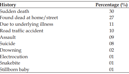
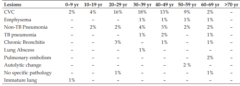
Ditribution of cases based on the clinical history
Description: No description available.
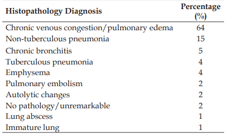
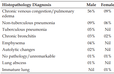
Distribution of cases based on histopathology diagnosis
Description: No description available.
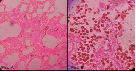
Photomicrographs show lung with features of congestion and presence of heart failure cells (hemosiderin laden macrophages) within the alveoli. Fig. A [H&E, 100X], Fig. B [H&E, 400X]
Description: No description available.
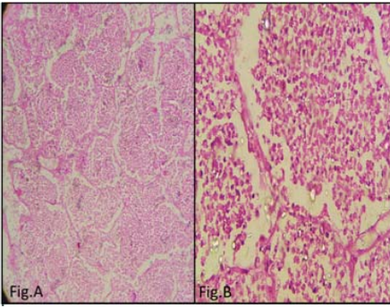
Photomicrographs show lung with features of lobar pneumonia. Fig. A [H&E, 100X], Fig. B [H&E, 400X]
Description: No description available.
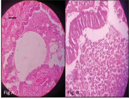
Photomicrographs show features of chronic bronchitis. (A & B) Bronchi with increased mucous glands (arrow) and presence of chronic inflammatory infiltrate within the bronchi. Fig. A [H&E, 100X], Fig. B [H&E, 400X]
Description: No description available.
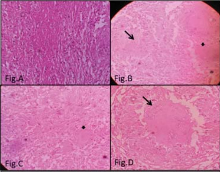
(A) Photomicrograph show caseous necrosis [H&E, 100X], (B & C) Photomicrographs show presence of granulomas with epithelioid cells (large arrow) and Langhans giant cell (arrow head) [H&E, 100X], (D) Photomicrograph show a well formed granuloma with epithelioid cells and central necrosis (arrow) [H&E, 400X]
Description: No description available.
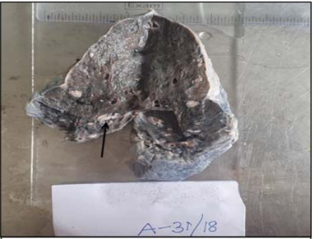
Shows gross picture of lung with the presence of multiple grey white nodules in the lower lobe (arrow), from a patient who was a known case of pulmonary tuberculosis
Description: No description available.
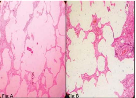
(A & B) Photomicrographs show lung with emphysematous changes [H&E, 100X]
Description: No description available.
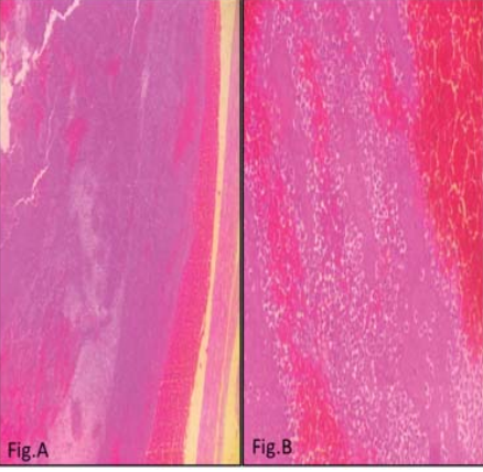
Photomicrograph of pulmonary emboli. (A) shows a pulmonary vessel with an embolus [H&E, 100X], (B) shows lines of zahn [H&E, 400X].