Full Text (PDF)
Indian Journal of Forensic Medicine and Pathology 19(3):p 335-345, July -Sept. 2026. | DOI: https://doi.org/10.21088/ijfmp.0974.3383.19326.8
Original Article
Histopathological Spectrum of Autopsy Cases: A One Year Retrospective Observational Study in a Tertiary Care Centre in Mumbai
Devanshi Shah, Manjusha Dhawle, Shweta Watane, Abhishek Muley, Ashwini Waso, Abhishikta Misra, Sushma Ramraje
Author Information
Licence:
Attribution-Non-commercial 4.0 International (CC BY-NC 4.0)This license enables reusers to distribute, remix, adapt, and build upon the material in any medium or format for noncommercial purposes only, and only so long as attribution is given to the creator.
Indian Journal of Forensic Medicine and Pathology 19(3):p 335-345, July -Sept. 2026. | DOI: https://doi.org/10.21088/ijfmp.0974.3383.19326.8
How Cite This Article:
Manjusha Dhawle, Shweta Watane, Abhishek Muley, et al. Histopathological Spectrum of Autopsy Cases: A One Year Retrospective Observational Study in a Tertiary Care Centre in Mumbai. Indian J Forensic Med Pathol. 2026; 19(3): 335-345.Timeline
Received : April 17, 2026
Accepted : June 22, 2026
Published : September 30, 2026
Abstract
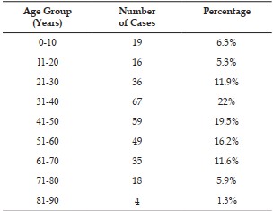
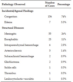
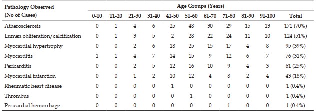
Introduction: Autopsy continues to play a significant role in understanding disease processes and identifying clinically silent pathological lesions that may remain undetected during life. Several of these lesions remain clinically silent throughout life and are detected only during autopsy. The present study was undertaken to evaluate incidental neoplastic and non-neoplastic lesions detected on histopathological examination of medicolegal autopsies. Aims and Objectives: 1. To evaluate histopathological lesions detected during autopsy and assess their association with the cause of death. 2. To classify the cause of death according to the various organ systems. 3. To classify the cause of death according to age and gender. Materials and Methods: A retrospective observational study was carried out in the Department of Pathology at a tertiary care centre over a period of one year from February 2024 to January 2025. The study included all the medicolegal autopsy specimens and viscera received for histopathological examination during the study period. A total of 303 cases were evaluated. Representative tissue sections from organs including brain, heart, lungs, liver, spleen, and kidneys were routinely processed and stained using hematoxylin and eosin stain. Microscopic examination was performed to identify pathological lesions and correlate findings with the clinical and autopsy details. Results: The present study consisted of a series of 303 autopsy cases from the Department of Pathology in a tertiary care hospital conducted over a period of one year. The internal organs of a total of 303 autopsies were sent for histopathological examination. The most commonly involved age group was 30-40 years, with a male preponderance. Neoplastic lesions were identified in 4.95% of cases. Conclusion: This study has contributed valuable insights to the spectrum of both neoplastic and non-neoplastic lesions, encompassing incidental as well as clinically significant findings. Routine autopsy histopathology facilitates identification of clinically unsuspected lesions and contributes valuable information regarding disease patterns and cause of death.
References
- 1. Sulegaon R., Kulkarni D., Chulki S. Medicolegal autopsies: Interesting and incidental findings. Int J Forensic Sci Pathol. 2015; 3(8): 156–160.
- 2. Sarvaiya A.N., Panjvani S.I., Shah N.R., Shah C.K. Incidental and interesting histopathological findings in medicolegal autopsies. Int J Sci Res. 2014; 3(1): 372–374.
- 3. Nadesan K. The importance of the medicolegal autopsy. Malays J Pathol. 1997; 19(2): 105–109.
- 4. Ludwig J. Handbook of autopsy practice. 3rd ed.
- 5. Molina D.K., Wood L.E., Frost R.E. Is routine histopathologic examination beneficial in all medicolegal autopsies? Am J Forensic Med Pathol. 2007; 28(1): 1–3.
- 6. Patel S., Rajalakshmi B.R., Manjunath G.V. Histopathologic findings in autopsies with emphasis on interesting and incidental findings: a pathologist’s perspective. J Clin Diagn Res. 2016; 10(11): EC08–EC12. doi:10.7860/JCDR/2016/22008.8835
- 7. Pathak A, Mangal HM. Histopathology examination in medico-legal autopsy: pros and cons. J Indian Acad Forensic Med. 2010;32:128.
- 8. Patel C.B., Patel K., Bhagat V.M., Shah P. Pattern of histopathological lesions in lung autopsy. Int J Res Med Sci. 2018; 6: 279–283.
- 9. Panchal M.G., Sonwane R.G. Histopathological study of MLC and autopsy cases in our hospital. Indian J Forensic Med Pathol. 2019; 12(2): 106–112.
- 10. Arunalatha P., Sangeetha A., Sumitra Devi N.R. Spectrum of histopathological findings in autopsies: highlighting interesting and incidental findings. Int J Curr Med Appl Sci. 2017; 15(2): 61–66.
- 11. Kandy N.C., Pai MR, Philipose R. Role of histopathology in autopsy study: an audit. SAS J Med. 2015; 1(1): 7–15.
- 12. Garg S., Hasija S., Sharma P., Kalhan S., Saini N., Khan A. Histopathological analysis of prevalence of various heart diseases: an autopsy study. Int J Res Med Sci. 2018; 6: 1414– 1418.
- 13. Sandhu V.K., Puri A, Singh N. Histomorphological spectrum of renal lesions in an autopsy study. Ann Pathol Lab Med. 2017; 4(4): A410–A414. doi:10.21276/APALM.1455
- 14. Bal M.S., Singh S.P., Bodal V.K., Oberoi S.S., Surinder K. Pathological findings in liver autopsy. J Indian Acad Forensic Med. 2004; 26(2): 55–57.
- 15. Jadhav D.S., Deshmukh S.S., Keswani A.S., et al. Study of histopathological examination in autopsy specimens at a tertiary care center in Maharashtra. Indian J Forensic Med Pathol. 2024; 17(4): 271–276.
- 16. Adelusola K.A., Osasan S.A., Afolabi O.A. Histopathological study and audit of the spleen in Nigerians. Afr J Health Sci. 2007; 14: 195–200.
- 17. Mohan B.P., Sivaraman L., Vijayan S., Das A.G. Spectrum of incidental tumors in autopsy specimens: A histopathological analysis of 797 cases. Indian J Health Sci Biomed Res. 2023; 16(2): 292–297.
Data Sharing Statement
There are no additional data available. All raw data and code are available upon request.
Funding
This research received no funding.
Author Contributions
All authors contributed significantly to the work and approve its publication.
Ethics Declaration
This article does not involve any human or animal subjects, and therefore does not require ethics approval.
Acknowledgements
We would like to express our gratitude to the patients, their families, and all those who have contributed to this study.
Conflicts of Interest
No conflicts of interest in this work.
About this article

Cite this article
Manjusha Dhawle, Shweta Watane, Abhishek Muley, et al. Histopathological Spectrum of Autopsy Cases: A One Year Retrospective Observational Study in a Tertiary Care Centre in Mumbai. Indian J Forensic Med Pathol. 2026; 19(3): 335-345.
Licence:
Attribution-Non-commercial 4.0 International (CC BY-NC 4.0)This license enables reusers to distribute, remix, adapt, and build upon the material in any medium or format for noncommercial purposes only, and only so long as attribution is given to the creator.
| Received | Accepted | Published |
|---|---|---|
| April 17, 2026 | June 22, 2026 | September 30, 2026 |
DOI: https://doi.org/10.21088/ijfmp.0974.3383.19326.8
Keywords
AutopsyHistopathologyLesionsMedicolegalSearch for Similar Articles
Similar Articles
- Diatom Analysis in Thermophilic water bodies: A Study on Hot Water Springs of Ma...
- Morphometric Study of Supra-meatal Triangle and Mastoid process in the Adult dry...
- Forensic Differentiation of Manner of Death in Railway Fatalities: A FourCase Se...
- Understanding assault Patterns: A Cross-sectional study of Cases at Chri, Kelamb...
- Assessing Response Awareness to a Snake Bite in Population of Pune Region
Article Level Metrics
Last UpdatedSaturday 17 October 2026, 01:02:12 (IST)
16152
Accesses
37
3203
00
Citations
NA
NA
NA
Download citation
Article Keywords
Keyword Highlighting
Highlight selected keywords in the article text.
Timeline
| Received | April 17, 2026 |
| Accepted | June 22, 2026 |
| Published | September 30, 2026 |
licence
Attribution-Non-commercial 4.0 International (CC BY-NC 4.0)
This license enables reusers to distribute, remix, adapt, and build upon the material in any medium or format for noncommercial purposes only, and only so long as attribution is given to the creator.