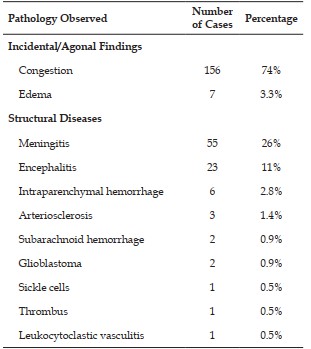
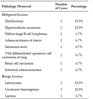
Description: Brain-Distribution of Cases According to Pathology observed
Heading
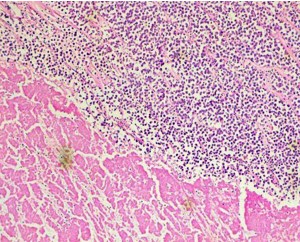
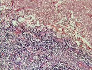
Description: Photomicrograph showing meningitis (H&E, 20x): Inflammatory infiltrate comprising of lymphocytes and plasma cells within meninges
Heading
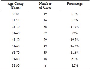
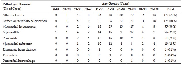
Description: Heart-Distribution of cases according to age and pathology observed
Heading
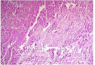
Description: Photomicrograph showing Old healed infarct (H&E, 20x): Myocardial hypertrophy, myocytolysis and collagenous scar replacing necrosed myocardial tissue
Heading
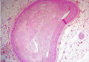
Description: Photomicrograph showing Atherosclerosis of coronary artery (H&E, 40x): Showing cholesterol in the fibrous cap with occlusion of lumen
Heading
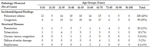
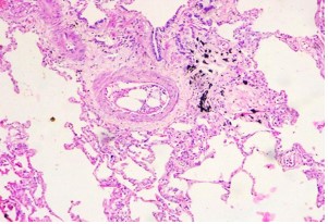
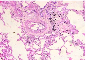
Description: Lung - Distribution of Cases According to Pathology observed
Heading
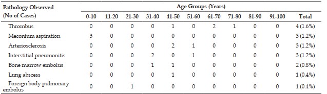
Description: Lung - Distribution of Cases According to Pathology observed
Heading
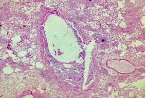
Description: Photomicrograph showing Bone marrow embolus (H&E, 20x): Heterogeneous mixture of hematopoietic elements and adipose tissue in pulmonary vessels
Heading
Description: Photomicrograph showing Diffuse alveolar damage (H&E, 20x): Pulmonary edema with congestion and interstitial inflammatory infiltrate with hyaline membrane formation in alveolar septa
Heading
Description: Photomicrograph showing Foreign body pulmonary embolus (IV drug abuse) (H&E, 20x): Foreign body granuloma comprising of foreign body giant cells and lymphocytes surrounding foreign body pale blue rod like material around perivascular areas and basophilic material within vessels
Heading
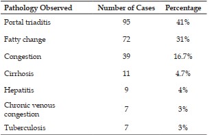
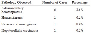
Description: Liver-Distribution of Cases According to Pathology observed
Heading
Description: Liver-Distribution of Cases According to Pathology observed
Heading
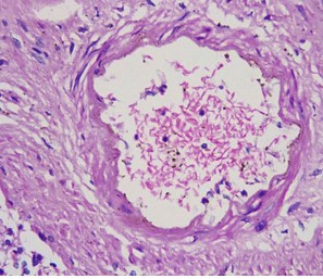
Description: Photomicrograph showing Sickle shaped RBCs (H&E, 40x): Sickle shaped RBCs in lumen of hepatic vessels
Heading
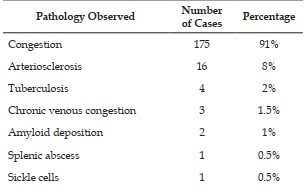
Description: Spleen-Distribution of Cases According to Pathology observed
Heading
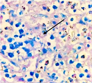
Description: Photomicrograph showing Tuberculosis spleen (ZN staining, 40x): Acid fast bacilli in spleen
Heading
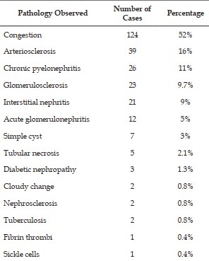
Description: Kidney-Distribution of Cases According to Pathology observed
Heading
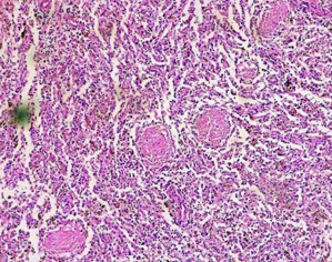
Description: Photomicrograph showing Amyloidosis kidney (H&E, 20x): Extracellular deposition of amyloid in glomeruli
Heading
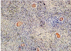
Description: Photomicrograph showing Amyloidosis kidney (Congo red stain, 20x)
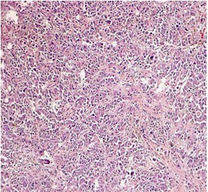
Description: Photomicrograph showing Hepatocellular carcinoma (H&E, 20x): Malignant hepatocytes arranged in cords and trabeculae
Heading
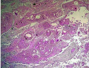
Description: Photomicrograph showing Well differentiated squamous cell carcinoma lung (H&E, 20x): Large, polygonal tumour cells in sheets with keratin pearl formation
Heading
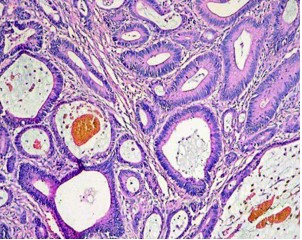
Description: Photomicrograph showing Adenocarcinoma of intestine (H&E, 20x): Glandular arrangement of tumour cells showing elongated, hyperchromatic nuclei
Heading
Description: Photomicrograph showing Seminoma testis (H&E, 20x): Sheets of pale tumour cells arranged in poorly demarcated lobules seperated by delicate septa with adjoining area of necrosis
Heading
Description: Photomicrograph showing Glioblastoma (H&E, 20x): Highly cellular astrocytic tumour with marked pleomorphism, nuclear atypia, brisk mitosis and palisading necrosis