Full Text (PDF)
Indian Journal of Forensic Medicine and Pathology 11(2):p 89-94, April-June 2018. | DOI: 10.21088/ijfmp.0974.3383.11218.8
Original Article
Histopathological Study of Cardiac Lesions in Medicolegal Autopsies
Medha P. , Kulkarni, Kalpana R. Sulhyan, Nayan A. Ramteerthakar
Author Information
Licence:
Attribution-Non-commercial 4.0 International (CC BY-NC 4.0)This license enables reusers to distribute, remix, adapt, and build upon the material in any medium or format for noncommercial purposes only, and only so long as attribution is given to the creator.
Indian Journal of Forensic Medicine and Pathology 11(2):p 89-94, April-June 2018. | DOI: 10.21088/ijfmp.0974.3383.11218.8
How Cite This Article:
Timeline
Received : January 31, 2018
Accepted : February 26, 2018
Published : March 30, 2018
Abstract
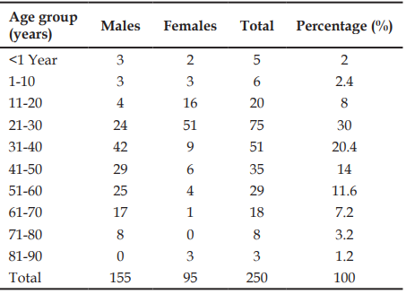
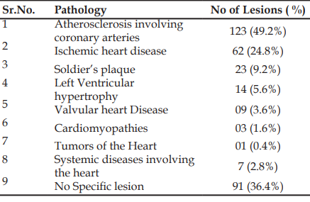
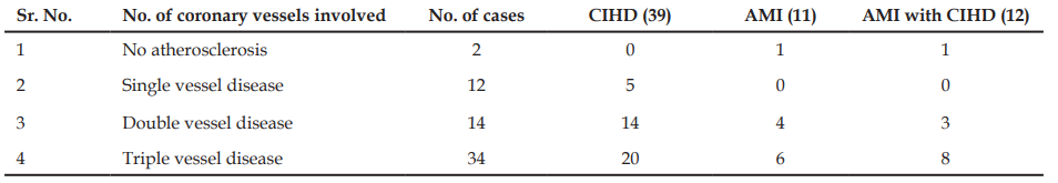
Context: Cardiovascular diseases are an important cause of morbidity and mortality in both developing and developed countries. The prevalence of coronary artery disease has doubled in Indians during the past three to four decades. Hence, the study was undertaken to evaluate the prevalence of various heart diseases in this region of western Maharashtra. Aim: The aim was to assess the pattern and prevalence of atherosclerosis involving the coronary arteries and to study the gross and microscopic features of various lesions affecting the valves, myocardium, endocardium and pericardium. Design: This was a prospective analytical study. Material and methods: The study included 250 complete heart specimens of medicolegal autopsy cases received from August 2012 to July 2014. Thorough gross examination of atria, ventricles and valves was done followed by examination of the coronary arteries and representative tissue bits were submitted for microscopy. Results: Atherosclerosis was the most common lesion involving 123 cases with 48 cases of triple vessel disease. Ischemic heart disease was seen in 62 cases, 60 being associated with atherosclerosis. Fourteen cases of left ventricular hypertrophy, nine cases of valvular heart disease, three cases each of cardiomyopathy and disseminated intravascular coagulation, two cases of acute leukemia and one case each of papillary fibroelastoma, amyloidosis, ventricular septal defect and sickle cell anaemia were observed. Conclusion: Present study emphasizes the role of atherosclerosis in ischemic heart disease. Amyloidosis and rare tumors like papillary fibroelastoma of the heart can be accurately diagnosed on histopathology underscoring the usefullness of an autopsy based study.
References
- 1. Schoen FJ, Mitchell RN. The Heart. In: Kumar V, Abbas AK, Fausto N, editors. Robbins and Cotran pathologic basis of disease. 8th ed. Philadelphia: Saunders Elsevier; 2010. p. 530.
- 2. Burke A, Tavora F. Practical cardiovascular pathology. 1st ed. Philadelphia: Lippincott Williams & Wilkins; 2011. p. 3-10.
- 3. Stary HC, Chandler AB, Dinsmore RE, Fuster V, Glagov S, Insull W, et al. A definition of advanced types of atherosclerotic lesions and a histological classification of atherosclerosis. A report from the Committee on Vascular Lesions of the Council on Arteriosclerosis, American Heart Association. Circulation. 1995;92(5):1355-74.
- 4. Marwah N, Sethi B, Gupta S, Duhan A, Singh S, Sen R. Histomorphological spectrum of various cardiac changes in sudden death: an autopsy study. Iran J Pathol. 2011;6(4):179-86.
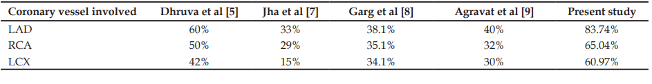
- 5. Dhruva GA, Agravat AH, Sanghvi HK. Atherosclerosis of coronary arteries as predisposing factor in myocardial infarction: an autopsy study. Online J Health Allied Sci. 2012;11(3):1-4.
- 6. Shiladaria P, Chauhan G, Parghi B, Goswami A, Suri S. Coronary atherosclerosis and myocardial infarction: an autopsy study. Natl J Integr Res Med. 2013;4(4):106-8.
- 7. Jha BM, Naik D, Agarwal A, Jana S, Patel M. Incidence of atherosclerosis in different coronary arteries and its relation with myocardial infarction: a randomized study in 300 autopsy hearts in tertiary care hospital. Int J Med Sci Public Health. 2013;2(4):836-9.
- 8. Garg M, Aggarwal AD, Kataria SP. Coronary atherosclerosis and myocardial infarction: an autopsy study. J Indian Acad Forensic Med. 2011;33(1):39-42.
- 9. Agravat A, Dhruva G, Babaria K, Rathod K. Coronary artery disease on autopsy: a five years clinicopathological study (November – 2007 to October – 2012). Int J Biomed Adv Res. 2013;4(2):105-11.
- 10. Travis WD, Brambilla E, Muller-Hermelink HK, Harris CC, editors. Pathology and genetics of tumours of the lung, pleura, thymus and heart. World Health Organization classification of tumours. Lyon: IARC Press; 2004. p. 313-5.
Data Sharing Statement
There are no additional data available. All raw data and code are available upon request.
Funding
This research received no funding.
Author Contributions
Whether all authors contributed significantly to the work and approve its publication.
Ethics Declaration
This article does not involve any human or animal subjects, and therefore does not require ethics approval.
Acknowledgements
We would like to express our gratitude to the patients, their families, and all those who have contributed to this study.
Conflicts of Interest
The authors report no conflicts of interest in this work.
About this article
Cite this article
Licence:
Attribution-Non-commercial 4.0 International (CC BY-NC 4.0)This license enables reusers to distribute, remix, adapt, and build upon the material in any medium or format for noncommercial purposes only, and only so long as attribution is given to the creator.
| Received | Accepted | Published |
|---|---|---|
| January 31, 2018 | February 26, 2018 | March 30, 2018 |
DOI: 10.21088/ijfmp.0974.3383.11218.8
Keywords
AtherosclerosisIschemic Heart DiseaseValvular Heart DiseaseSearch for Similar Articles
Similar Articles
- Pesticide Contamination in Indian Agricultural and Residential Areas: A Compara...
- Forensic Age Estimation using CBCT-Derived Mandibular Morphometrics: A Comparat...
- A 2 Years Retrospective Study of the Spectrum of Poisoning in a Tertiary Care C...
- Knowledge and Attitude of MBBS Students Regarding Post Mortem Examination: A Cr...
- Sudden Death in a 7-year-old Child Due to Neoplasm: A Case Report
Article Level Metrics
Last UpdatedWednesday 08 July 2026, 07:41:09 (IST)
7723
Accesses
3
2137
00
Citations
NA
NA
NA
Download citation
Article Keywords
Keyword Highlighting
Highlight selected keywords in the article text.
Timeline
| Received | January 31, 2018 |
| Accepted | February 26, 2018 |
| Published | March 30, 2018 |
licence
Attribution-Non-commercial 4.0 International (CC BY-NC 4.0)
This license enables reusers to distribute, remix, adapt, and build upon the material in any medium or format for noncommercial purposes only, and only so long as attribution is given to the creator.