Full Text (PDF)
Indian Journal of Forensic Medicine and Pathology 19(2):p 133-144, April - June 2026. | DOI: https://doi.org/10.21088/ijfmp.0974.3383.19226.3
Original Article
Forensic Age Estimation using CBCT-Derived Mandibular Morphometrics: A Comparative Study of Regression and Machine-Learning Models
Kamala K.A., Asheerbad Swain, Suraiya Khan, Anish Nelson, Rahul Tiwari, Heena Dixit Tiwari, Manish Sharma
Author Information
Licence:
Attribution-Non-commercial 4.0 International (CC BY-NC 4.0)This license enables reusers to distribute, remix, adapt, and build upon the material in any medium or format for noncommercial purposes only, and only so long as attribution is given to the creator.
Indian Journal of Forensic Medicine and Pathology 19(2):p 133-144, April - June 2026. | DOI: https://doi.org/10.21088/ijfmp.0974.3383.19226.3
How Cite This Article:
Asheerbad Swain, Kamala K.A., Suraiya Khan, et al. Forensic Age Estimation Using CBCT-Derived Mandibular Morphometrics: A Comparative Study of Regression and Machine-Learning Models. Indian J Forensic Med Pathol. 2026; 19(2): 133-144.Timeline
Received : December 29, 2025
Accepted : March 06, 2026
Published : June 30, 2026
Abstract
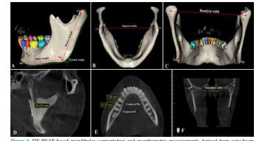
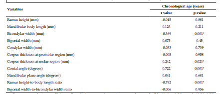
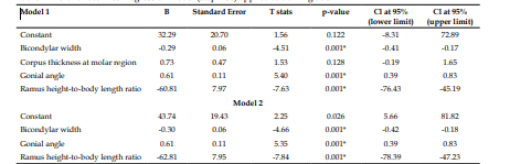
Background: Accurate age estimation in adolescents and adults remains challenging in forensic practice once dental development is complete. Cone-beam computed tomography (CBCT) enables three-dimensional evaluation of skeletal structures and may improve age estimation without additional radiation exposure. Aim: To develop and internally validate a CBCT-based multivariate regression model for chronological age estimation using mandibular morphometrics and to compare its performance with a machine-learning approach. Materials and Methods: This retrospective study analyzed 150 CBCT scans of individuals aged 10–70 years. Mandibles were segmented using ITK-SNAP software, and standardized three-dimensional morphometric measurements were obtained. The dataset was randomly divided into a training set (n testing set (n = 45). Pearson’s correlation analysis and stepwise multivariate linear regression were used to develop the regression model. A Random Forest regression model was trained for comparison. Model performance was assessed using the coefficient of determination (R²), root mean square error (RMSE), mean absolute error (MAE), and mean absolute percentage error (MAPE). Agreement between predicted and chronological age was evaluated using Bland–Altman analysis and intraclass correlation coefficient (ICC). Results: Chronological age showed a strong positive correlation with the gonial angle and a strong negative correlation with the ramus height–to–body length ratio. The final regression model retained gonial angle, bicondylar width, and ramus height–to–body length ratio as significant predictors. In the testing dataset, the regression model demonstrated excellent predictive accuracy (R² = 0.881; RMSE = 6.09 years; MAE = 5.89 years), minimal bias (−0.49 years), and excellent agreement (ICC = 0.90). The Random Forest model showed reasonable performance but did not outperform the regression model. Conclusion: CBCT-derived mandibular morphometrics enable accurate, noninvasive forensic age estimation. The regressi on model demonstrated superior reliability and interpretability compared with machine-learning, supporting its clinical and medico-legal applicability.= 105) and a
References
- 1. Ubelaker D.H., Khosrowshahi H. Estimation of age in forensic anthropology: historical perspective and recent methodological advances. Forensic Sci Res. 2019; 4(1): 1-9. doi: 10.1080/20961790.2018.1549711. .
- 2. Gabriel D.B., Southard K.A., Qian F, Marshall S.D., Franciscus R.G., Southard T.E. Cervical vertebrae maturation method: poor reproducibility. Am J Orthod Dentofacial Orthop. 2009; 136(4): 478.e1-7; discussion 478-80. doi: 10.1016/j.ajodo.2007.08.028.
- 3. Venkatesh E., Elluru S.V. Cone beam computed tomography: basics and applications in dentistry. J Istanb Univ Fac Dent. 2017; 51(3 Suppl 1): S102-S121. doi: 10.17096/jiufd.00289.
- 4. Motawei S.M., Helaly A.M., Aboelmaaty W.M., Elmahdy K., Shabka O.A., Liu H. Length of the ramus of the mandible as an indicator of chronological age and sex: a study in a group of Egyptians. Forensic Sci Int. 2020; 2 doi: 10.1016/j.fsir.2020.100066.
- 5. Dogan M.E., Turkoglu B.N., Şengul I. A Morphometric Evaluation of the Mandibular Condyle, Coronoid Process, and Gonial Angle: Age and Gender Differences in CBCT Imaging. Diagnostics (Basel). 2025; 15(12): 1459. doi: 10.3390/diagnostics15121459.
- 6. Arthanari A., Senthilkumar A., Ramalingam K., Prathap L., Ravindran V. Exploring Age and Gender Identification Through Mandibular Parameters Using Orthopantomography: An Observational Study. Cureus. 2024; 16(3): e55788. doi: 10.7759/cureus.55788.
- 7. Puspa Larasati L.D., Kurniawan A., Chusida A., Rizky B.N., Marini M.I., Arisa Putri Q.O., et al. Geometric morphometric analysis of mandibular morphology for age classification in Indonesian adolescents and adults. J Oral Biol Craniofac Res. 2025; 15(6): 1518-1525. doi: 10.1016/j.jobcr.2025.09.011.
- 8. Green S.B. How many subjects does it take to do a regression analysis. Multivariate Behav Res. 1991; 26(3): 499-510. doi: 10.1207/ s15327906mbr2603_7
- 9. Vila-Blanco N., Varas-Quintana P., Tomás I., Carreira M.J. A systematic overview of dental methods for age assessment in living individuals: from traditional to artificial intelligence-based approaches. Int J Legal Med. 2023; 137(4): 1117-1146. doi: 10.1007/s00414- 023-02960-z.
- 10. Izard G. The gonio-mandibular angle in dentofacial orthopedia. Int J Orthodontia. 1927; 13: 578. 10.1016/S0099-6963(27)90110-0
- 11. Ohm E., Silness J. Size of the mandibular jaw angle related to age, tooth retention and gender. J Oral Rehabil. 1999; 26(11): 883-91. doi: 10.1046/j.1365-2842.1999.00464.x.
- 12. Leversha J., McKeough G., Myrteza A, Skjellrup-Wakefiled H., Welsh J., Sholapurkar A. Age and gender correlation of gonial angle, ramus height and bigonial width in dentate subjects in a dental school in Far North Queensland. J Clin Exp Dent. 2016; 8(1): e49-54. doi: 10.4317/jced.52683.
- 13. Bakan A., Kervancıoğlu P., Bahşi İ., Yalçın E.D. Comparison of the Gonial Angle With Age and Gender Using Cone-Beam Computed Tomography Images. Cureus. 2022; 14(5): e24997. doi: 10.7759/cureus.24997.
- 14. Mitteroecker P., Schaefer K. Thirty years of geometric morphometrics: Achievements, challenges, and the ongoing quest for biological meaningfulness. Am J Biol Anthropol. 2022; 178 Suppl 74(Suppl 74): 181-210. doi: 10.1002/ ajpa.24531.
- 15. Al-Ghurabi Z.H., Al-Bahrani Z.M. Cone Beam Computed Tomography Evaluation of the Morphological Variation and Width in Mandibular Condyle. J Craniofac Surg. 2021; 32(5): e479-e481. doi: 10.1097/ SCS.0000000000007465.
- 16. Üzel A., Kuran A., Baysal O., Seki U., Sinanoglu E.A. Age estimation by radiomics analysis of mandibular condylar cone beam computed Asheerbad Swain, Kamala K.A., Suraiya Khan, et al. Forensic Age Estimation Using CBCT-Derived Mandibular Morphometrics: A Comparative Study of Regression and Machine-Learning Models.144 Indian Journal of Forensic Medicine and Pathology IJFMP/Volume 19 Number 2 April–June 2026 tomography images. Leg Med (Tokyo). 2025; 72: 102560. doi: 10.1016/j.legalmed.2024.102560.
- 17. Valladares Neto J., Estrela C., Bueno M.R., Guedes O.A., Porto O.C.L., Pécora J.D. Mandibular condyle dimensional changes in subjects from 3 to 20 years of age using ConeBeam Computed Tomography: A preliminary study. Dental Press J Orthod. 2010; 15: 172-81. https://www.scielo.br/j/dpjo/a/XzKjjZxRL mvnxWDTpnKG5fJ/?format=pdf&lang=en
- 18. Singh R., Singh H., Aggarwal N., Sehgal G., Chandel S., Kumar N. Sexual Dimorphism in Mandibular Ramus Morphometry: A Population-Specific Analysis Using Orthopantomograms in the Lucknow Region. Cureus. 2025; 17(3): e80515. doi: 10.7759/ cureus.80515.
- 19. Kvaal S.I., Kolltveit K.M., Thomsen I.O., Solheim T. Age estimation of adults from dental radiographs. Forensic Sci Int. 1995; 74(3): 175-85. doi: 10.1016/0379-0738(95)01760-g.
- 20. Cameriere R., Ferrante L., Cingolani M. Variations in pulp/tooth area ratio as an indicator of age: a preliminary study. J Forensic Sci. 2004; 49(2): 317-9. https://pubmed.ncbi. nlm.nih.gov/15027553/
- 21. Vodanovic M., Sribar A., Tomljenovic F., Milosevic D., Subasic M. Age Estimation Based On Orthopantomograms Using Artificial Intelligence. Int Dent J. 2025; 75: 104186. 10.1016/j.identj.2025.104186 22. Yushkevich P.A., Piven J., Hazlett H.C., Smith R.G., Ho S., Gee J.C., Gerig G. User-guided 3D active contour segmentation of anatomical structures: significantly improved efficiency and reliability. Neuroimage. 2006; 31(3): 1116- 28. doi: 10.1016/j.neuroimage.2006.01.015.
- 23. Yushkevich P.A., Yang Gao, Gerig G. ITKSNAP: An interactive tool for semi-automatic segmentation of multi-modality biomedical images. Annu Int Conf IEEE Eng Med Biol Soc. 2016; 2016: 3342-3345. doi: 10.1109/ EMBC.2016.7591443.
Data Sharing Statement
There are no additional data available. All raw data and code are available upon request.
Funding
This research received no funding.
Author Contributions
All authors contributed significantly to the work and approve its publication.
Ethics Declaration
This article does not involve any human or animal subjects, and therefore does not require ethics approval.
Acknowledgements
We would like to express our gratitude to the patients, their families, and all those who have contributed to this study.
Conflicts of Interest
No conflicts of interest in this work.
About this article

Cite this article
Asheerbad Swain, Kamala K.A., Suraiya Khan, et al. Forensic Age Estimation Using CBCT-Derived Mandibular Morphometrics: A Comparative Study of Regression and Machine-Learning Models. Indian J Forensic Med Pathol. 2026; 19(2): 133-144.
Licence:
Attribution-Non-commercial 4.0 International (CC BY-NC 4.0)This license enables reusers to distribute, remix, adapt, and build upon the material in any medium or format for noncommercial purposes only, and only so long as attribution is given to the creator.
| Received | Accepted | Published |
|---|---|---|
| December 29, 2025 | March 06, 2026 | June 30, 2026 |
DOI: https://doi.org/10.21088/ijfmp.0974.3383.19226.3
Keywords
Age •EstimationForensicMandibularMorphologySearch for Similar Articles
Similar Articles
- Diatom Analysis in Thermophilic water bodies: A Study on Hot Water Springs of Ma...
- Morphometric Study of Supra-meatal Triangle and Mastoid process in the Adult dry...
- Histopathological Spectrum of Autopsy Cases: A One Year Retrospective Observatio...
- Forensic Differentiation of Manner of Death in Railway Fatalities: A FourCase Se...
- Understanding assault Patterns: A Cross-sectional study of Cases at Chri, Kelamb...
Article Level Metrics
Last UpdatedWednesday 21 October 2026, 14:19:14 (IST)
16514
Accesses
49
3226
00
Citations
NA
NA
NA
Download citation
Article Keywords
Keyword Highlighting
Highlight selected keywords in the article text.
Timeline
| Received | December 29, 2025 |
| Accepted | March 06, 2026 |
| Published | June 30, 2026 |
licence
Attribution-Non-commercial 4.0 International (CC BY-NC 4.0)
This license enables reusers to distribute, remix, adapt, and build upon the material in any medium or format for noncommercial purposes only, and only so long as attribution is given to the creator.