Full Text (PDF)
Indian Journal of Forensic Medicine and Pathology 17(3):p 185-189, July- September 2024. | DOI: https://doi.org/10.21088/ijfmp.0974.3383.17324.5
Original Article
Determination of Sex Using Mastoid Process
Vishnu Prasad, Thumma Amar, Mr. Vedarethinam, Vijayakumar Nair G, Shilpa T Patil
Author Information
Licence:
Attribution-Non-commercial 4.0 International (CC BY-NC 4.0)This license enables reusers to distribute, remix, adapt, and build upon the material in any medium or format for noncommercial purposes only, and only so long as attribution is given to the creator.
Indian Journal of Forensic Medicine and Pathology 17(3):p 185-189, July- September 2024. | DOI: https://doi.org/10.21088/ijfmp.0974.3383.17324.5
How Cite This Article:
Prasad V, Amar T, Vedarethinam, et al. Determination of Sex Using Mastoid Process. Indian J Forensic Med Pathol. 2024;17(3):185-189.Timeline
Received : July 02, 2024
Accepted : August 31, 2024
Published : September 25, 2024
Abstract
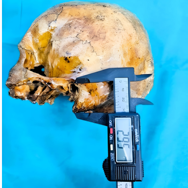
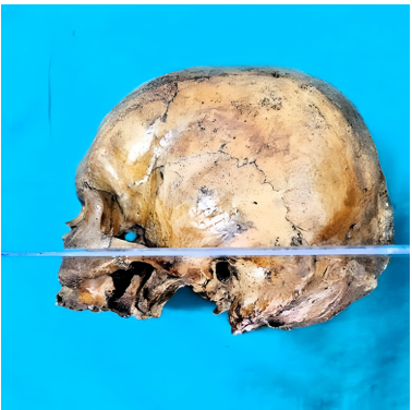
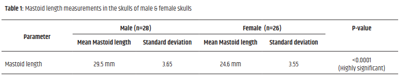
Background: Anthropometric studies of bones play a crucial role in establishing identification, especially in fragmented skeletal remains. Mastoid process is acone shaped bony protuberance extending from mastoid region of temporal bone of the skull. It is usually undamaged, because of its unique and secured position. The present study is aimed at evaluating the usefulness of the mastoid length in gender identification. Material and Methods: 54 dried, and unharmed skulls of identified sex were employed in the study. Out of which 28 were male, 26 were female. The mastoid process length was measured on either side by using a Vernier calliper from Frankfurt’s plane. Results: In male skulls, the mean mastoid length was 29.5 ± 3.65 mm, while in female skulls, it was 24.6 ± 3.55 mm. A statistical analysis indicated that the mastoid length for determining sex has a highly significant p value of less than 0.0001. Conclusion: The mean mastoid length is significantly less in females than in males. Therefore, it can be considered as a sexual dimorphic feature and is useful in determining sex from fragmented skull.
References
- 1. Krogman WM. The human skeleton in forensic medicine. Springfield, IL: Charles C Thomas, 1962.
- 2. Bilge Y, Kedici PS, Alakoç YD, Ülküer KÜ, and İlkyaz YY. The identification of a dismembered human body: A multidisciplinary approach. Forensic Sci Int 2003; 137(2-3): 141-146.
- 3. Whall J and Henke W. The pars petrosa as diagnostic aid in multivariate biometric sex determination of creamated human remains Z MorpholAnthropol1980, 70: 258-68.
- 4. Kalmey JK, and Rathbun TA. Sex determination by discriminant function analysis of petrous portion of temporal bone. J Forensic Sci 1996; 41: 865-867.
- 5. Paiva LAS, Segre M. Sexing the human skull through the mastoid process. Rev Hosp ClnFac Med Sao Paulo 2003;58(1):15–20.
- 6. Nagaoka T, Shizushima A, Sawada J, Tomo S, Hoshino K, Sato H, et al. Sex determination using mastoid process measurements: standards for Japanese human skeletons of the medieval and early modern periods. Anthropol Sci 2008; 116(2):105–13.
- 7. Passey J, Mishra SR, Singh R, Sushobhna K, Singh S, Sinha P. Sex determination using mastoid process. Asian Journal of Medical Sciences. 2015 Jun 1; 6(6):93–5.
- 8. Hoshi H. Sex difference in the shape of the mastoid process in norma occipitalis and its importance to the sex determination of the human skull. Okajma’s Folia Ana Japonica 1962; 38:309-17.
- 9. Keen JA. Sex differences in skulls. American Journal of Physical Anthropology 1950; 8: 65-79.
- 10. Giles E., and Elliot O. Sex determination by discriminant function analysis of crania. American Journal of Physical Anthropology 1963; 21: 53-68.
- 11. Sumati Patnaik VVG, and Phatak A. Determination of sex from mastoid process by discriminant function analysis. J Anat So India 2010; 59(2):222-8.
- 12. Song HW, Lin ZQ, Jia JT. Sex diagnosis of Chinese skulls using multiple stepwise discrinant function analysis. Forensic Sci Int 1992; 54(2);135-40.
- 13. Patil KR, Mody RN. Determination of sex by discriminant function analysis ans stature by regression analysis a lateral cephalometric study. Forensic Sci In 2005; 147(2- 3):175-80.
Data Sharing Statement
There are no additional data available. All raw data and code are available upon request.
Funding
This research received no funding.
Author Contributions
All authors contributed significantly to the work and approve its publication.
Ethics Declaration
This article does not involve any human or animal subjects, and therefore does not require ethics approval.
Acknowledgements
We would like to express our gratitude to the patients, their families, and all those who have contributed to this study.
Conflicts of Interest
No conflicts of interest in this work.
About this article
Cite this article
Prasad V, Amar T, Vedarethinam, et al. Determination of Sex Using Mastoid Process. Indian J Forensic Med Pathol. 2024;17(3):185-189.
Licence:
Attribution-Non-commercial 4.0 International (CC BY-NC 4.0)This license enables reusers to distribute, remix, adapt, and build upon the material in any medium or format for noncommercial purposes only, and only so long as attribution is given to the creator.
| Received | Accepted | Published |
|---|---|---|
| July 02, 2024 | August 31, 2024 | September 25, 2024 |
DOI: https://doi.org/10.21088/ijfmp.0974.3383.17324.5
Keywords
AnthropometrySexual dimorphismMastoid lengthSearch for Similar Articles
Similar Articles
- Pesticide Contamination in Indian Agricultural and Residential Areas: A Compara...
- Forensic Age Estimation using CBCT-Derived Mandibular Morphometrics: A Comparat...
- A 2 Years Retrospective Study of the Spectrum of Poisoning in a Tertiary Care C...
- Knowledge and Attitude of MBBS Students Regarding Post Mortem Examination: A Cr...
- Sudden Death in a 7-year-old Child Due to Neoplasm: A Case Report
Article Level Metrics
Last UpdatedWednesday 08 July 2026, 05:34:35 (IST)
7722
Accesses
24
2137
00
Citations
NA
NA
NA
Download citation
Article Keywords
Keyword Highlighting
Highlight selected keywords in the article text.
Timeline
| Received | July 02, 2024 |
| Accepted | August 31, 2024 |
| Published | September 25, 2024 |
licence
Attribution-Non-commercial 4.0 International (CC BY-NC 4.0)
This license enables reusers to distribute, remix, adapt, and build upon the material in any medium or format for noncommercial purposes only, and only so long as attribution is given to the creator.