Full Text (PDF)
Indian Journal of Forensic Medicine and Pathology 15(4):p 281-287, October-December 2022. | DOI: https://doi.org/10.21088/ijfmp.0974.3383.15422.11
Original Article
Determination of Gender Divergences of Maxillary Sinuses Measurements in Computed Tomographic Scans of Kerala Population
Tina Sharma, Vyshnav R, Priyakanksha Mishra
Author Information
Licence:
Attribution-Non-commercial 4.0 International (CC BY-NC 4.0)This license enables reusers to distribute, remix, adapt, and build upon the material in any medium or format for noncommercial purposes only, and only so long as attribution is given to the creator
Indian Journal of Forensic Medicine and Pathology 15(4):p 281-287, October-December 2022. | DOI: https://doi.org/10.21088/ijfmp.0974.3383.15422.11
How Cite This Article:
Vyshnav R, Sharma T, Mishra P. Determination of gender divergences of maxillary sinuses measurements in computed tomographic scans of Kerala population. Indian J Forensic Med Pathol. 2022;15(4):281-287.Timeline
Received : July 20, 2022
Accepted : October 28, 2022
Published : December 30, 2022
Abstract
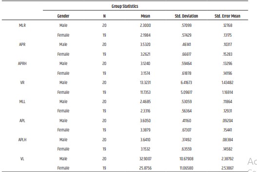
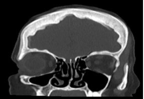
The challenging forensic procedure of corpse identification is required by law authorities and social norms. It is very crucial to compare the post-mortem and antemortem medical information in cases where the bodies are severely damaged and beyond recognition. However, conventional identification techniques might not have been effective, especially though there have been substantial post-mortem alterations. In unidentifiable skeletons, gender has long been determined from either the skull, pelvic or long bones with an epiphysis and a metaphysis. In the present study, an attempt has been made to determine gender divergences from axial and coronal computed tomographic scans (CT) of the maxillary sinus in the Kerala population. A total of 40 individuals including 20 males and 20 females, visiting the Outpatient Department of the Koyilli Hospital, Kannur were included as the study subjects. The dimensions of the right and left maxillary sinuses of 40 subjects from plain CT were measured using Radiant DICOM software. Statistical analysis was completed with an independent student t-test and general descriptive analysis with SPSS software. Gender determination using height, length, and the width of the maxillary sinus on both sides showed statistically insignificant results but in the case of left volume and left the anterior-posterior height of the maxillary sinus the p-value was found to be significant at 95% of confidence level. So, the present study concludes that the left volume of the maxillary sinus and left anteroposterior height showed statistically significant results which can be considered in determining and screening corpses on the bases of maxillary sinus computed tomographic records.
References
- 1. Sidhu, R., Chandra, S., Devi, P., Taneja, N., Sah, K., & Kaur, N. Forensic importance of maxillary sinus in gender determination: A morphometric analysis from Western Uttar Pradesh, India. European Journal of General Dentistry.2014: 3(01), 53-56.
- 2. Fernandes, C. L. Forensic ethnic identification of crania: the role of the maxillary sinus—a new approach. The American journal of forensic medicine and pathology. 2014: 25(4), 302-313.
- 3. Sahlstrand-Johnson, P., Jannert, M., Strömbeck, A., & Abul-Kasim, K. Computed tomography measurements of different dimensions of maxillary and frontal sinuses. BMC medical imaging. 1992; 11(1), 1-7.
- 4. Reichs, K. J. Quantified comparison of frontal sinus patterns by means of computed tomography. Forensic science international. 1993: 61(2-3), 141-168.
- 5. Zhen, S., Ma, X., Lu, B., Ming, S., Lin, K., Zhao, L., Zhou, W. Supercapacitor electrodes based on furan-EDOT copolymers via electropolymerization. International Journal of Electrochemical Science. 9(12), 2014: 7518-7527.
- 6. Teke, H. Y., Duran, S., Canturk, N., & Canturk, G. Determination of gender by measuring the size of the maxillary sinuses in computerized tomography scans. Surgical and radiologic anatomy. 2007: 29(1), 9-13.
- 7. Wind, J., & Zonneveld, F. W. Computed Tomography of an AustrMopithecus Skull (Mrs Pies): A New Technique. 1989:7518-7527.
- 8. Uthman, A. T., Al-Rawi, N. H., & AlTimimi, J. F. Evaluation of foramen magnum in gender determination using helical CT scanning. Dentomaxillofacial Radiology. 2012: 41(3), 197-202.
- 9. Ariji, Y., Ariji, E., Yoshiura, K., & Kanda, S. Computed tomographic indices for maxillary sinus size in comparison with the sinus volume. Dentomaxillofacial Radiology. 25(1), 19-24.
- 10. Sidhu, R., Chandra, S., Devi, P., Taneja, N., Sah, K., & Kaur, N. Forensic importance of maxillary sinus in gender determination: A morphometric analysis from Western Uttar Pradesh, India. European Journal of General Dentistry. 2014: 3(01), 53-56.
- 11. Teke, H. Y., Duran, S., Canturk, N., & Canturk, G. Determination of gender by measuring the size of the maxillary sinuses in computerized tomography scans. Surgical and radiologic anatomy. 2007:29(1), 9-13.
- 12. Kanchan, T., & Rastogi, P. Sex determination from hand dimensions of North and South Indians. Journal of forensic sciences. 2009: 54(3), 546-550.
- 13. Uthman, A. T., Al‐Rawi, N. H., Al‐ Naaimi, A. S., & Al‐Timimi, J. F. Evaluation of maxillary sinus dimensions in gender determination using helical CT scanning. Journal of forensic sciences. 2011: 56(2), 403- 408.
- 14. Eshak, G. A., Ahmed, H. M., & Gawad, E. A. A. Gender determination from hand bones length and volume using multidetector computed tomography: a study in Egyptian people. Journal of forensic and legal medicine. 2011: 18(6), 246-252.
- 15. Amin, M. F., & Hassan, E. I. Sex identification in Egyptian population using Multidetector Computed Tomography of the maxillary sinus. Journal of forensic and legal medicine. 2012: 19(2), 65- 69.
- 16. Giacomini, G., Pavan, A. L. M., Altemani, J. M. C., Duarte, S. B., Fortaleza, C. M. C. B., Miranda, J. R. D. A., & De Pina, D. R. Computed tomography-based volumetric tool for standardized measurement of the maxillary sinus. PloS one. 2018: 13(1), e0190770.
- 17. Riepert, T., Ulmcke, D., Schweden, F., & Nafe, B. Identification of unknown dead bodies by X-ray image comparison of the skull using the X-ray simulation program Foxsis. Forensic science international. 2001: 117(1-2), 89-98.
- 18. Quatrehomme, G., Fronty, P., Sapanet, M., Grévin, G., Bailet, P., & Ollier, A. Identification by frontal sinus pattern in forensic anthropology. Forensic science international. 1996: 83(2), 147-153.
- 19. Pirner, S., Tingelhoff, K., Wagner, I., Westphal, R., Rilk, M., Wahl, F. M., Eichhorn, K. W. (2009). CT-based manual segmentation and evaluation of paranasal sinuses. European archives of oto-rhinolaryngology. 2009: 266(4), 507-518.
- 20. Ekizoglu, O., Inci, E., Hocaoglu, E., Sayin, I., Kayhan, F. T., & Can, I. O. (2014). The use of maxillary sinus dimensions in gender determination: a thin-slice multidetector computed tomography assisted morphometric study. Journal of Craniofacial Surgery. 2014: 25(3), 957-960.
Data Sharing Statement
There are no additional data available. All raw data and code are available upon request.
Funding
This research received no funding.
Author Contributions
All authors contributed significantly to the work and approve its publication.
Ethics Declaration
This article does not involve any human or animal subjects, and therefore does not require ethics approval.
Acknowledgements
We would like to express our gratitude to the patients, their families, and all those who have contributed to this study.
Conflicts of Interest
No conflicts of interest in this work.
About this article
Cite this article
Vyshnav R, Sharma T, Mishra P. Determination of gender divergences of maxillary sinuses measurements in computed tomographic scans of Kerala population. Indian J Forensic Med Pathol. 2022;15(4):281-287.
Licence:
Attribution-Non-commercial 4.0 International (CC BY-NC 4.0)This license enables reusers to distribute, remix, adapt, and build upon the material in any medium or format for noncommercial purposes only, and only so long as attribution is given to the creator
| Received | Accepted | Published |
|---|---|---|
| July 20, 2022 | October 28, 2022 | December 30, 2022 |
DOI: https://doi.org/10.21088/ijfmp.0974.3383.15422.11
Keywords
Forensic ScienceForensic MedicineGender determinationForensic AnthropologyPostmortem IdentificationMaxillary Sinus.Forensic ScienceForensic MedicineGender determinationForensic AnthropologyPostmortem IdentificationMaxillary Sinus.Search for Similar Articles
Similar Articles
- Pesticide Contamination in Indian Agricultural and Residential Areas: A Compara...
- Forensic Age Estimation using CBCT-Derived Mandibular Morphometrics: A Comparat...
- A 2 Years Retrospective Study of the Spectrum of Poisoning in a Tertiary Care C...
- Knowledge and Attitude of MBBS Students Regarding Post Mortem Examination: A Cr...
- Sudden Death in a 7-year-old Child Due to Neoplasm: A Case Report
Article Level Metrics
Last UpdatedWednesday 08 July 2026, 06:42:44 (IST)
7722
Accesses
16
2137
00
Citations
NA
NA
NA
Download citation
Article Keywords
Keyword Highlighting
Highlight selected keywords in the article text.
Timeline
| Received | July 20, 2022 |
| Accepted | October 28, 2022 |
| Published | December 30, 2022 |
licence
Attribution-Non-commercial 4.0 International (CC BY-NC 4.0)
This license enables reusers to distribute, remix, adapt, and build upon the material in any medium or format for noncommercial purposes only, and only so long as attribution is given to the creator