Full Text (PDF)
Indian Journal of Pathology: Research and Practice 11(1):p 19-26, January-March 2022. | DOI: https://doi.org/10.21088/ijprp.2278.148X.11122.2
Original Article
COVID-19 Associated Mucormycosis (CAM): A Clinico Histopathological Study of at A Tertiary Care Centre
Sunil V Jagtap, Swati S Jagtap, Atul Hulwan, Snigdha A Vartak, Divya Brahmbhatt, Shubhang Monpara
Author Information
Licence:
Attribution-Non-commercial 4.0 International (CC BY-NC 4.0)This license enables reusers to distribute, remix, adapt, and build upon the material in any medium or format for noncommercial purposes only, and only so long as attribution is given to the creator.
Indian Journal of Pathology: Research and Practice 11(1):p 19-26, January-March 2022. | DOI: https://doi.org/10.21088/ijprp.2278.148X.11122.2
How Cite This Article:
Sunil V Jagtap, Swati S Jagtap, Atul Hulwan, et. al./COVID-19 Associated Mucormycosis (CAM): A Clinico Histopathological Study of at A Tertiary Care Centre/J Pathol Res Pract 2022;11(1):19–26.Timeline
Received : December 31, 2024
Accepted : February 01, 2022
Published : March 30, 2022
Abstract
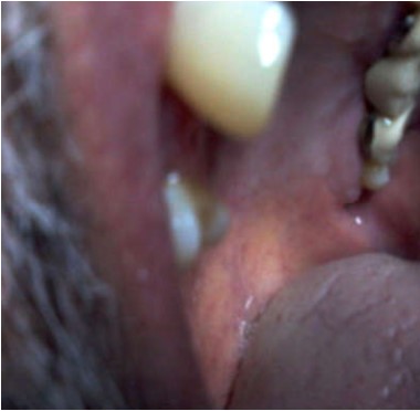
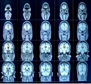
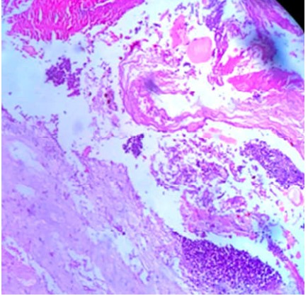
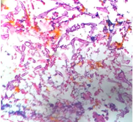
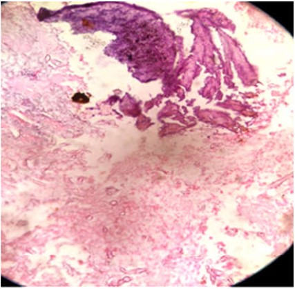
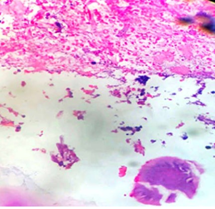
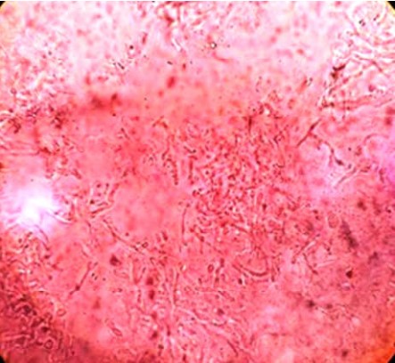
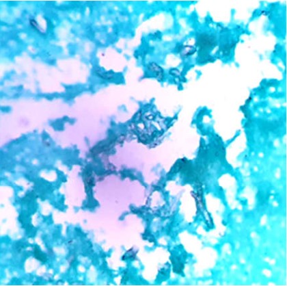
During the current pandemic of COVID-19 there are increased cases with super infections by mucormycosis. This study is performed with an aim to evaluate clinico histomorphological features of COVID-19 associated mucormycosis(CAM). Material and Methods: A total of 3365 COVID 19 positive cases were reported during the last 8 months. A total of 494 patients with COVID 19 were admitted for >24 hours during this study period. Among these 13 cases of mucormycosis were diagnosed. All cases were positive for Reverse transcriptase polymerase chain reaction RT PCR assay. The detail clinical examination and required investigations were included. The operative details and histopathological gross and microscopic findings were noted. The characteristics type of inflammation, necrosis, fungal hyphae, presence of granulomatous reaction, angioinvasion and perineural invasion,bony tissue pathology were noted. The special stain like PAS, Grocott Gomori's methenaminesilver stainwere done. The data was analysed. Results: The age range of patients was from 10 to 65 year. There was a male preponderance. The 6 (46%) patients had pre existing diabetes mellitus, 3 (23%) patients had received steroids during the treatment, 1 patient hadcoexisting HIV/AIDS infection, 3 (23%) patients had history of hypertension. The radio imaging revealed soft tissue swelling in the preseptal, malar, premaxillary and retrobulbar regions with paranasal sinusitis. Nasal involvement was seen in all 13 cases (100%), orbital involvement in 5 cases (37%) and cerebral involvement in 3 cases (23%). On histomorphological study characteristic fungal hyphae of mucormycosis were seen in all cases. The acute or chronic inflammation, necrosis were seen in all cases. The granulomatous reaction was seen in 23%, angioinvasion in 46% and perineural invasion in 15% cases. One case revealed co-exisisting HIV/AIDS and another with actinomycosis co-infection. Conclusions: In COVID 19 patients Mucormycosis is one of the important associated infection within this pandemic. This increase in incidence is related to various risk factors being systemic preexisting disease, IV infection, diabetics, use of steroid and immunosuppression etc.The mucormycosis patients are treated by early identification, monitoring risk factors, followed by proper surgical intervention and administration of the appropriate antifungal medication. This histopathological study will be of importance in early diagnosis and management of patients with COVID-19 and would help improve outcomes in world pandemic time. We are presenting these casesof CAM for its clinical, radio imaging, and histopathological findings.
References
- 1. Coronaviridae Study Group of the International Committee on Taxonomy of Viruses. The species Severe acute respiratory syndrome-related coronavirus: classifying 2019-nCoV and naming it SARS-CoV-2. Nat Microbiol.2020;5, 536–544.
- 2. World Health Organization. Novel coronavirus China. 2020 (https://www.who.int/csr/don/12-january-2020-novel-coronavirus-china/en/in). Emergency Situational Updates.Weekly epidemiological update on COVID-19 - 27 July 2021.Edition 50,27 July 2021.
- 3. Kubin CJ, McConville TH, Dietz D, et al. Characterization of Bacterial and Fungal Infections in Hospitalized Patients With Coronavirus Disease 2019 and Factors Associated With Health CareAssociated Infections. Open Forum Infect Dis. 2021;8(6):of201.
- 4. Woon H. Chong, Biplab K. Saha, Ananthakrishnan Ramani, Amit Chopra. State-of-the-art review of secondary pulmonary infections in patients with COVID-19 pneumonia. Infection 49:4, 591-605.
- 5. Eucker J., Sezer O., Graf B., Possinger K. Mucormycoses. Mycoses. 2001;44(7):253–260
- 6. Skiada A, Pavleas I, Drogari-Apiranthitou M. Epidemiology and Diagnosis of Mucormycosis: An Update. J Fungi (Basel). 2020;6(4):265.
- 7. Adhikari S.P., Meng S., Wu Y.J., Mao Y.P et al.Epidemiology, causes, clinical manifestation and diagnosis, prevention and control of coronavirus disease (COVID-19)during the early outbreak period: a scoping review. Infect. Dis. Poverty. 2020;17(9):29.
- 8. Global Action Fund for Fungal Infections (GAFFI). Doctors around the world call for rapid response to deadly mucormycosis (the so-called “black fungus”) found in COVID patients in India. Available at https://bit.ly/352wj9F
- 9. Garg D, Muthu V, Sehgal IS, Ramachandran R, Kaur H, Bhalla A, Puri GD, Chakrabarti A, Agarwal R. Coronavirus Disease (Covid-19) Associated Mucormycosis (CAM): Case Report and Systematic Review of Literature. Mycopathologia. 2021;186(2):289-298.
- 10. Shiv LalSoni, Kamal Kajal, L. N. Yaddanapudi, Pankaj Malhotra, GoverdhanDuttPuri, Ashish Bhalla, et al. Demographic and clinical prolfile of patients with COVID-19 at a tertiary care hospital in north India. Indian J Med Res 2021;153:115-125.
- 11. Mehta S, Pandey A. Rhino-Orbital Mucormycosis Associated With COVID-19. Cureus 2020;12(9).
- 12. Rawson TM, Moore LSP, Zhu N, et al.: Bacterial and fungal co-infection in individuals with coronavirus: a rapid review to support COVID-19 antimicrobial prescribing. Clin Infect Dis. 2020,3;71(9):2459-68.
- 13. Y.P. Talmi, A. Goldschmied,Reouven, M. Bakon, I. Barshack, M. Wolf, Z. Horowitz, et al.Rhinoorbital and rhino-orbito-cerebral mucormycosis. Otolaryngol. Head Neck Surg., 2002;127,1 : 22-31.
- 14. Jagtap S V, Jagtap S S, Nagar V , Varshney K , Invasive mucormycosis in post COVID-19 infection: Case report with review. IP Arch Cytol Histopathol Res 2021;6(2):135-139.
- 15. Prakash H., Chakrabarti A. Global epidemiology of mucormycosis. J Fungi. 2019;5:26.
- 16. Patel A, Kaur H, Xess I, Michael JS, Savio J, Rudramurthy S, et al. A multicentre observational study on the epidemiology, risk factors, management and outcomes of mucormycosis in India. Clin Microbiol Infect. 2020;26:944.e9–15.
- 17. International Diabetes Federation . (2020). Accessed: July 7, 2020: https://idf.org/our-network/regionsmembers/south-east-asia/members/94-india.html.
- 18. Hoang K., Abdo T., Reinersman J.M., Lu R., Higuita N.I.A. A case of invasive pulmonary mucormycosis resulting from short courses of corticosteroids in a well-controlled diabetic patient. Med Mycol Case Rep. 2020;29(1):22–24
- 19. RajeevSoman, Ayesha Sunavala. Post COVID-19 Mucormycosis – from the Frying Pan into the Fire. JAPI Jan 2021;69:46.
- 20. Chowdhary A, Tarai B, Singh A, Sharma A: Multidrug-resistant Candida auris infections in critically ill coronavirus disease patients, India, April-July 2020. Emerg Infect Dis. 2020, 26:11. 10.
- 21. Roden MM, Zaoutis TE, Buchanan WL, et al. Epidemiology and outcome of zygomycosis: a review of 929 reported cases. Clin Infect Dis 2005; 41: 634–53.
- 22. Saldanha M,Reddy, R.& Vincent, M.J. Title of the Article: Paranasal Mucormycosis in COVID-19 Patient. Indian J Otolaryngol Head Neck Surg ,2021 ;22:1-4.
- 23. Singh AK, Singh R, Joshi SR, Misra A. Mucormycosis in COVID-19: A systematic review of cases reported worldwide and in India [published online ahead of print, 2021 May 21]. Diabetes Metab Syndr. 2021;15(4):102146.
- 24. Wang X, Sahu KK, Cerny J. Coagulopathy, endothelial dysfunction, thrombotic microan -giopathy and complement activation: potential role of complement system inhibition in COVID-19. J Thromb Thrombolysis. 2021 ;51(3):657-662.
- 25. Tiwari NR, Phatak S, Sharma VR, Agarwal SK. COVID-19 and thrombotic microangiopathies. Thromb Res. 2021 ;202:191-198.
- 26. Sravani T, Uppin SG, Uppin MS, Sundaram C. Rhinocerebralmucormycosis: Pathology revisited with emphasis on perineural spread. Neurol India. 2014;62:383-6.
- 27. Kandpal H, Aneesh MK, Seith A, Sharma S. Symptomatic perineural extension of fungal sinusitis in an immune competent person: Imaging features. Singapore Med J. 2008;49:171–4.
- 28. Gamba JL, Woodruff WW, Djang WT, Yeates AE. Craniofacial mucormycosis: Assessment with CT. Radiology. 1986;160:207–7.
- 29. Jagtap SV, Hulwan A, Vartak S et.al. Co-infection of mucormycosis and actinomycosis in COVID-19 infection. Int J Health Sci Res. 2021; 11(8): 127-130.
- 30. Cornely OA, Alastruey-Izquierdo A, Arenz D, Chen SCA, Dannaoui E, Hochhegger B, et al. Mucormycosis ECMM MSG Global GuidelineWriting Group. Global guideline for the diagnosis and management of mucormycosis: an initiative of the European Confederation of Medical Mycology in cooperation with the Mycoses Study Group Education and Research Consortium. Lancet Infect Dis. 2019 ;19(12):e405-e421.
Data Sharing Statement
There are no additional data available. All raw data and code are available upon request.
Funding
This research received no funding.
Author Contributions
All authors contributed significantly to the work and approve its publication.
Ethics Declaration
This article does not involve any human or animal subjects, and therefore does not require ethics approval.
Acknowledgements
We would like to express our gratitude to the patients, their families, and all those who have contributed to this study.
Conflicts of Interest
No conflicts of interest in this work.
About this article

Cite this article
Sunil V Jagtap, Swati S Jagtap, Atul Hulwan, et. al./COVID-19 Associated Mucormycosis (CAM): A Clinico Histopathological Study of at A Tertiary Care Centre/J Pathol Res Pract 2022;11(1):19–26.
Licence:
Attribution-Non-commercial 4.0 International (CC BY-NC 4.0)This license enables reusers to distribute, remix, adapt, and build upon the material in any medium or format for noncommercial purposes only, and only so long as attribution is given to the creator.
| Received | Accepted | Published |
|---|---|---|
| December 31, 2024 | February 01, 2022 | March 30, 2022 |
DOI: https://doi.org/10.21088/ijprp.2278.148X.11122.2
Keywords
COVID-19MucormycosisFungal infectionsRT-PCR TestSearch for Similar Articles
Similar Articles
- Clinico-Pathological and Immunohistochemical Study of Primary CNS Tumors in a Te...
- The Role of SGLT2 Inhibitors in Cardiovascular Protection Beyond Diabetes: A Co...
- Epidermal Inclusion Cyst Presenting as 4th Ventricle Space Occupying Lesion: A R...
- Silent Retroperitoneal Seminoma Arising from an Occult Undescended Testis: Diagn...
- Scapular Metastasis of Oral Squamous Cell Carcinoma: A Case Report of an Unusual...
Article Level Metrics
Last UpdatedSaturday 17 October 2026, 10:30:26 (IST)
4177
Accesses
10
671
00
Citations
NA
NA
NA
Download citation
Article Keywords
Keyword Highlighting
Highlight selected keywords in the article text.
Timeline
| Received | December 31, 2024 |
| Accepted | February 01, 2022 |
| Published | March 30, 2022 |
licence
Attribution-Non-commercial 4.0 International (CC BY-NC 4.0)
This license enables reusers to distribute, remix, adapt, and build upon the material in any medium or format for noncommercial purposes only, and only so long as attribution is given to the creator.