Full Text (PDF)
Indian Journal of Forensic Medicine and Pathology 17(2):p 101-107, January – June 2024. | DOI: https://doi.org/10.21088/ijfmp.0974.3383.17224.5
Original Article
Cause and Manner of Death in Medicolegal Autopsy of Lung: A Tertiary Care Centre Study from North India
Dezy Singh, R. C. Tiwari, Ashish Bhute, Ravi P Meshram, Bhawana Mittal, Arvind Kumar
Author Information
Licence:
Attribution-Non-commercial 4.0 International (CC BY-NC 4.0)This license enables reusers to distribute, remix, adapt, and build upon the material in any medium or format for noncommercial purposes only, and only so long as attribution is given to the creator.
Indian Journal of Forensic Medicine and Pathology 17(2):p 101-107, January – June 2024. | DOI: https://doi.org/10.21088/ijfmp.0974.3383.17224.5
How Cite This Article:
Singh D, Tiwari RC, Bhute A, et al. Cause and Manner of Death in Medicolegal Autopsy of Lung: A Tertiary Care Centre Study from North India. Indian J Forensic Med Pathol. 2024;17(2):101-107.Timeline
Received : February 01, 2024
Accepted : June 20, 2024
Published : June 30, 2024
Abstract
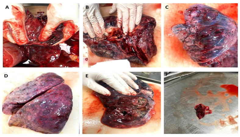
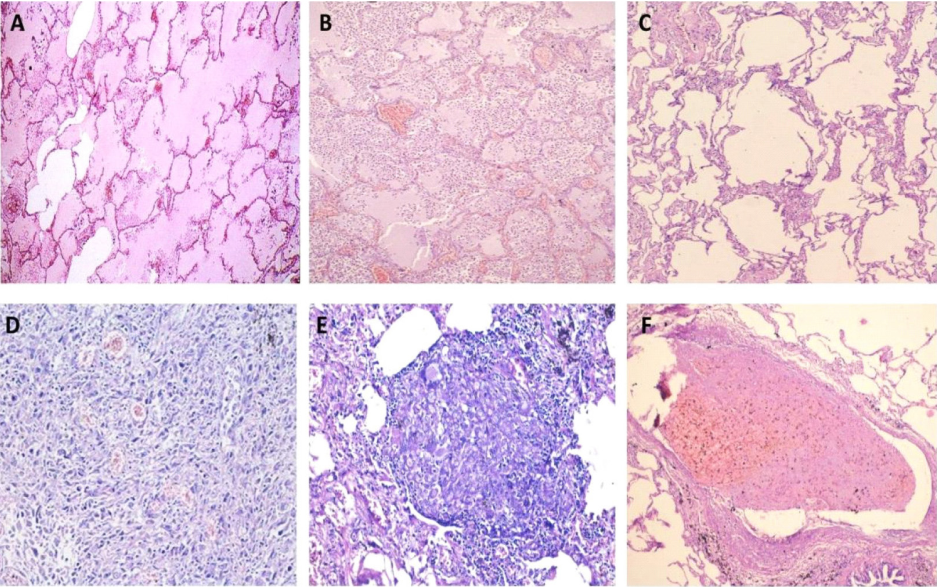
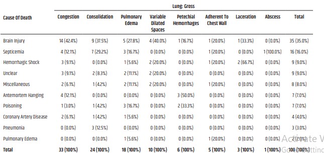
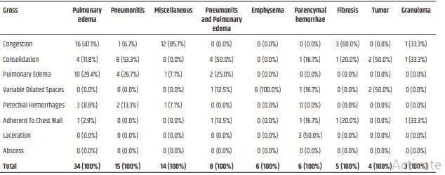
Context: Medicolegal autopsy results are determined by the pathological examination hence there is a need to check and consolidate all the factors that contribute in the interpretation of histology finding. Lung is vital organ in contributing the certain histologic pattern that can unfold the cause and manner of death. Aims: To note the role of gross and histopathological examination in ascertaining the cause and manner of death in lung autopsy. Setting and Design: Study was conducted in department of Pathology with Forensic department collaboration over the period of one year. It’s a prospective observational study including 100 medicolegal autopsy cases taken. Material and Method: Gross examination followed by histopathological examination on hemotyxilin and eosin stained slide of lung tissue were done. Special stains were used wherever required and findings noted. All variable were correlated using Chi-squared test, Fisher’s Exact and Kruskal Wallis and strength of association with Kendall’s Tau, Chi-squared test, Cramer’s V statistical tools. Results: Majority of cases were in between 20-39 yrs with male predominence. Most common gross finding was congestion followed by consolidation. In histology pulmonary edeme followed by pneumonitis were noted. Most frequent cause of death was brain injury followed by septicemia and manner of death was accidental followed by natural. There was significant association observed between histopathology and age as well as in between gross finding with cause and manner of death. Conclusion: Gross finding coupled with microscopy can link in the detection of cause and manner of death.
References
- 1. Vijaykumar L. Suicide and its prevention: The urgent need in India. Indian J Psychiatry2007; 49: 81-4.
- 2. Udayashankar SK, Shashikala P, Kavita GU, Pruthvi D. Histomorphological pattern of lung in medicolegal autopsies. Internat J Sci Res. 2015; 4:1937–9.
- 3. Singh D, Tiwari RC, Kumar A, Bhute AR, Meshram RP, Dikshit M, et al. A Comprehensive Review of Pathological Examination in Forensic Medicine: Past, Present, and Future. Cureus. 2022 Mar 1;14(3):e22740. doi: 10.7759/ cureus.22740.
- 4. Narayan Reddy KS, Murty OP. 33rd ed. Jaypee Brothers; 2014. The Essentials of Forensic Medicine and Toxicology.
- 5. Todorović MS, Mitrović S, Aleksandrić B, Mladjenović N, Matejić S. Association of pulmonary histopathological findings with toxicological findings in forensic autopsies of illicit drug users. Vojnosanit Pregl 2011 Aug;68(8):639- 42.
- 6. Jameson JL, Kasper D, Hauser S, Longo D, Fauci A, Loscalzo J. Harrison’s Principles of Internal Medicine. 19th ed. Vol. 2. Bhavnagar: Mc Graw Hill; 2015.
- 7. Minal G. Panchal, Rupali Giridhar Sonwane. Histopathological Study of MLC and Autopsy Cases in Our Hospital. Indian J Forensic Med Pathol. 2019;12(2):106-112.
- 8. P. Arunalatha, A. Sangeetha & Nalli. R. Sumitra Devi. Spectrum of Histopathological Findings in Autopsies - Highlighting the Interesting and Incidental Findings. International Journal of Current Medical and Applied Sciences. 2017 July;15(2):61-66.
- 9. Pulak Chakma et al. Indian Journal of Forensic and Community Medicine, July-September 2017;4(3):189-194 191.
- 10. Chauhan G, Agrawal M, Thakkar N, Parghi B. Spectrum of histopathological lesions in lung autopsy. J Res Med Den Sci 2015; 3(2):109-12.
Data Sharing Statement
There are no additional data available.
Funding
This research received no funding.
Author Contributions
All authors contributed significantly to the work and approve its publication.
Ethics Declaration
This article does not involve any human or animal subjects, and therefore does not require ethics approval.
Acknowledgements
Information not provide.
Conflicts of Interest
The authors report no conflicts of interest in this work.
About this article
Cite this article
Singh D, Tiwari RC, Bhute A, et al. Cause and Manner of Death in Medicolegal Autopsy of Lung: A Tertiary Care Centre Study from North India. Indian J Forensic Med Pathol. 2024;17(2):101-107.
Licence:
Attribution-Non-commercial 4.0 International (CC BY-NC 4.0)This license enables reusers to distribute, remix, adapt, and build upon the material in any medium or format for noncommercial purposes only, and only so long as attribution is given to the creator.
| Received | Accepted | Published |
|---|---|---|
| February 01, 2024 | June 20, 2024 | June 30, 2024 |
DOI: https://doi.org/10.21088/ijfmp.0974.3383.17224.5
Keywords
AutopsyCause of deathLungManner of death and pathologySearch for Similar Articles
Similar Articles
- Sudden Death in a 7-year-old Child Due to Neoplasm: A Case Report
- Bites of Burden: Snake Bite Envenoming in India, Regional Venom Differences, Tr...
- Forensic View of Suicide in Healthcare Workers
- Tracing Identity from Teeth to Technology: Artificial Intelligence in Forensic...
- Chemical Constituents in Latent Fingermarks as Indicators of Lifestyle using Gas...
Article Level Metrics
Last UpdatedWednesday 17 June 2026, 23:32:10 (IST)
7105
Accesses
6
2030
00
Citations
NA
NA
NA
Download citation
Article Keywords
Keyword Highlighting
Highlight selected keywords in the article text.
Timeline
| Received | February 01, 2024 |
| Accepted | June 20, 2024 |
| Published | June 30, 2024 |
licence
Attribution-Non-commercial 4.0 International (CC BY-NC 4.0)
This license enables reusers to distribute, remix, adapt, and build upon the material in any medium or format for noncommercial purposes only, and only so long as attribution is given to the creator.