Full Text (PDF)
Indian Journal of Forensic Medicine and Pathology 17(2):p 100-100, January – June 2024. | DOI: https://doi.org/10.21088/ijfmp.0974.3383.17224.4
Original Article
An Insight into Experiences of Forensic Expert and Pathologist on Prostrate Degeneration with Post Mortem Interval in Human Cadavers
Mopuri Venkateswarlu, C. Lakshmi Kalavathi, Ananth Rupesh Kattamreddy, Arun Ravula, Zunaid Ali Ahamed S
Author Information
Licence:
Attribution-Non-commercial 4.0 International (CC BY-NC 4.0)This license enables reusers to distribute, remix, adapt, and build upon the material in any medium or format for noncommercial purposes only, and only so long as attribution is given to the creator.
Indian Journal of Forensic Medicine and Pathology 17(2):p 100-100, January – June 2024. | DOI: https://doi.org/10.21088/ijfmp.0974.3383.17224.4
How Cite This Article:
Venkateswarlu M, Kalavathi CL, Kattamreddy AR, et al. An insight into experiences of Forensic expert and Pathologist on Prostrate Degeneration with Post Mortem interval in Human Cadavers. Indian J Forensic Med Pathol. 2024;17(2):95-100.Timeline
Received : November 17, 2023
Accepted : April 06, 2024
Published : June 30, 2024
Abstract
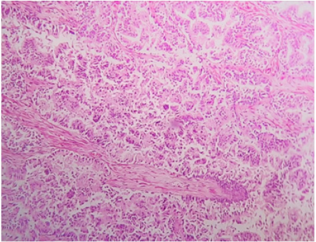
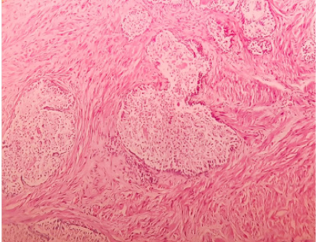
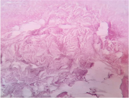
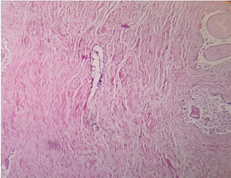
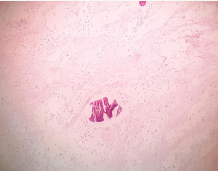
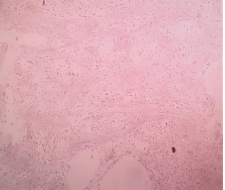
Background: Estimating postmortem interval is effective forensic tool for investigating time of death useful in criminology. Many gross, microscopic and molecular methods available for estimating postmortem interval. Human prostate is last soft tissue organ to degenerate in human cadavers and its histopathology in cadavers can be used for estimation of post mortem interval. Studies reported Histopathologies of different organs to estimate postmortem intervals for purpose of calculating time since death. In our study we tried to observe Post mortem interval estimation in human cadavers with Histopathological changes in Prostrate. Objectives: To estimate postmortem interval from Histopathological examination of prostate in human cadavers and identify Histopathological changes in human prostate in relation to time since death. Methodology: Prostate from cadavers registered for autopsy in our institute were examined grossly along with histopathology as per criteria laid down for sampling. Results: Histopathological sections from total of 36 human cadavers were studied. Changes like epithelial disruption of acini, nuclear changes, inflammatory cell collection in stroma, fatty degeneration and sequential necrotic changes were reported in relation to time since death. Earliest degeneration changes in prostrate acini began at 6 hours postmortem and changes in stroma began at 12 hours. First atrophic changes in acini began at 19 hours postmortem and continued to progress till 3 days after which identification of any glandular or stromal tissue became extremely difficult. Conclusion: Significant changes in Prostrate were documented between 6 hours and 72 hours postmortem. Changes in human prostate can be used for estimating postmortem interval.
References
- 1. Shrestha R, Kanchan T, Krishan K. Methods of Estimation of Time Since Death. [Updated 2020 Apr 29]. In: StatPearls [Internet]. Treasure Island (FL): StatPearls Publishing; 2020 Jan-. Available from: https://www.ncbi.nlm. nih.gov/books/NBK549867/.
- 2. Ceciliason, AS., Andersson, M.G., Nyberg, S. et al. Histological quantification of decomposed human livers: a potential aid for estimation of the post-mortem interval?. Int J Legal Med (2020). https:// doi.org/10.1007/s00414-020-02467-x.
- 3. Scrivano S, Sanavio M, Tozzo P, Caenazzo L. Analysis of RNA in the estimation of post-mortem interval: a review of current evidence. Int J Legal Med. 2019 Nov; 133(6):1629-1640.
- 4. Tomita Y, Nihira M, Ohno Y, Sato S. [Histological study of early postmortem changes in various organs: comparison of the paraffin embedding method and the epoxy resin embedding method]. Nihon HoigakuZasshi. 1999 Jun; 53(2):207-17. Japanese. PMID: 10536439.
- 5. Zhou C, Byard RW (2011) Factors and processes causing accelerated decomposition in human cadavers – an overview. J Forensic Legal Med 18:6–9. https://doi.org/10.1016/j. jflm.2010.10.003.
- 6. N Abdel Rahman Mahmoud, A Abdel Rahman Abdel Rahman Hassan, A Hassan Abdel Rahim, S Mostafa Mahmoud, O Hassan Nada, Molecular versus histopathological examination of the prostate gland in the estimation of post-mortem interval (an experimental study), QJM: An International Journal of Medicine, Volume 111, Issue suppl_1, December 2018, hcy200.054, https://doi. org/10.1093/qjmed/hcy200.05.
- 7. Mahmoud NA, Hassan AA, Abdel Rahim AH, Abdel Rahim AH, Suzan Mostafa Mahmoud SM, Nada OH. Molecular versus histopathological examination of the prostate gland in the estimation of post-mortem interval (an experimental study). Egypt. J. Forensic Sci. Appli. Toxicol. 2018;18(1):35-54.
- 8. Murty OP. Histopathology in forensic practice. Journal of Forensic Medicine and Toxicology. 2016;33(2):1-89.
- 9. Thakur R, Tiwari A. Estimation of time since death by histological examination of proximal convoluted tubule in human kidney. International Journal of Science & Healthcare Research. 2019; 4(4): 131-141.
- 10. Goncalves BF, de Campos SG, Costa CF, Scarano WR, Góes RM, Taboga SR. Key participants of the tumor microenvironment of the prostate: An approach of the structural dynamic of cellular elements and extracellular matrix components during epithelial–stromal transition. Actahistochemica. 2015 Jan 1;117(1):4-13.
Data Sharing Statement
There are no additional data available.
Funding
This research received no funding.
Author Contributions
All authors contributed significantly to the work and approve its publication.
Ethics Declaration
There is no ethical issue involved in the study and Post Mortem will be performed after obtaining necessary medico legal permissions and consents which also include organ observation and preservation in Human mortal specimens.
Acknowledgements
Information not provide.
Conflicts of Interest
The authors report no conflicts of interest in this work.
About this article
Cite this article
Venkateswarlu M, Kalavathi CL, Kattamreddy AR, et al. An insight into experiences of Forensic expert and Pathologist on Prostrate Degeneration with Post Mortem interval in Human Cadavers. Indian J Forensic Med Pathol. 2024;17(2):95-100.
Licence:
Attribution-Non-commercial 4.0 International (CC BY-NC 4.0)This license enables reusers to distribute, remix, adapt, and build upon the material in any medium or format for noncommercial purposes only, and only so long as attribution is given to the creator.
| Received | Accepted | Published |
|---|---|---|
| November 17, 2023 | April 06, 2024 | June 30, 2024 |
DOI: https://doi.org/10.21088/ijfmp.0974.3383.17224.4
Keywords
Forensic PathologyAutopsyHistologyProstateCriminalisticsSearch for Similar Articles
Similar Articles
- Analysis of Death Due to Pulmonary Embolism, a Case Series
- Suspected Adverse Event Following Immunization with Multisystem Inflammatory Res...
- Drone: A Smart Intelligent Framework Aiding Forensic Investigations
- Assessment of Knowledge on Sexual Assault Forensic Examination among Nurses in I...
- An Analysis of the Medico-Legal Aspects and Trends in Sexual Offense Cases at a...
Article Level Metrics
Last UpdatedThursday 09 July 2026, 08:55:39 (IST)
7829
Accesses
7
2153
00
Citations
NA
NA
NA
Download citation
Article Keywords
Keyword Highlighting
Highlight selected keywords in the article text.
Timeline
| Received | November 17, 2023 |
| Accepted | April 06, 2024 |
| Published | June 30, 2024 |
licence
Attribution-Non-commercial 4.0 International (CC BY-NC 4.0)
This license enables reusers to distribute, remix, adapt, and build upon the material in any medium or format for noncommercial purposes only, and only so long as attribution is given to the creator.