Full Text (PDF)
Indian Journal of Pathology: Research and Practice 12(3):p 95-103, July-September 2023. | DOI: https://doi.org/10.21088/ijprp.2278.148X.12323.2
Review Article
Benign Epithelial Inclusions in Lymph Nodes: A Diagnostic Challenge for the Pathologists
Mukta Pujani, Cherry Bansal, Meenu Pujani
Author Information
Licence:
Attribution-Non-commercial 4.0 International (CC BY-NC 4.0)This license enables reusers to distribute, remix, adapt, and build upon the material in any medium or format for noncommercial purposes only, and only so long as attribution is given to the creator
Indian Journal of Pathology: Research and Practice 12(3):p 95-103, July-September 2023. | DOI: https://doi.org/10.21088/ijprp.2278.148X.12323.2
How Cite This Article:
Mukta Pujani, Cherry Bansal, Meenu Pujani/Benign Epithelial Inclusions in Lymph Nodes: A Diagnostic Challenge for the Pathologists/Indian Journal of Pathology: Research and Practice 2023;12(3) 95–103.Timeline
Received : July 29, 2023
Accepted : September 02, 2023
Published : September 30, 2023
Abstract
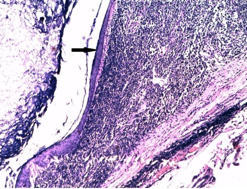
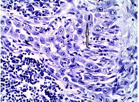
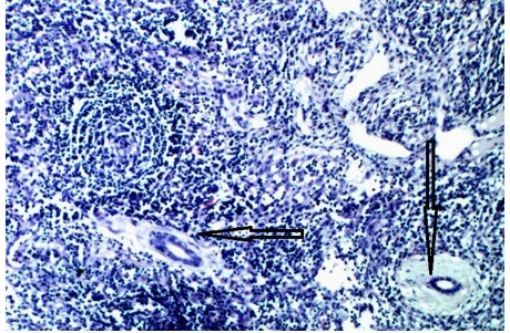
Benign inclusions are defined as foci of non neoplastic ectopic tissues in lymph nodes encompassing various types of tissues. Benign epithelial inclusions have been reported in lymph nodes of various anatomical locations including head and neck, mediastinum, axilla, peritoneal, pelvic and inguinal regions. Epithelial inclusions can be divided into epithelial, nevomelanocytic and decidual. Further subtypes based on the tissue of origin include salivary gland tissue, thyroid tissue, squamous epithelial cells, breast tissue, mesothelial cells etc. The nature of tissue in heterotopic inclusions varies according to the location of the lymph nodes. The accurate diagnosis of these benign epithelial inclusions is of utmost clinical importance to prevent misinterpretation of this entity as malignant and hence save the patient from overzealous and inappropriate treatment. Hence, great importance should be given to detailed morphological evaluation of lymph nodes with epithelial inclusions in reaching at an accurate diagnosis. The cytological criteria favouring benign epithelial inclusions over metastatic carcinoma include a lack of significant nuclear atypia, pleomorphism and hyperchromasia. It is very important to create awareness of this rare entity among pathologists to prevent misdiagnosis or overdiagnosis of a malignant lesion.
References
- 1. Ioachim HL, Medeiros LJ. Epithelial inclusions in lymph nodes. Ioachim HL, Medeiros LJ editors Ioachim's Lymph Node Pathology. Lippincott Williams & Wilkins, Fourth edition 2009:283-88.
- 2. Fregnani, JH, Latorre MR, Soares FA, Barros MD, Macéa JR. Estudo de inclusões epiteliais benignas nos linfonodos pélvicos de pacientes com carcinoma de colo de útero. AMHFCMSCSP 2004,49(2/3):61-5.
- 3. Spinardi JR, Goncalves IRD, La Falce TS, Fregnani JHTG, Barros MD, Macea JR. Benign inclusions in lymph nodes. Int J Morphol 2007; 25:625-9.
- 4. Wertheim E. Zur Frage der Radikaloperation beim Uteruskrebs. Arch Gynäkol, 61:627, 1900.
- 5. Maassen V, Hiller K. Glandular inclusions in lymph nodes: pattern of distribution and metaplastic transformation. Arch Gynecol Obstet. 1994;255(1):1-8.
- 6. Reich O, Tamussino K, Haas J, Winter R. Benign müllerian inclusions in pelvic and paraaortic lymph nodes. Gynecol Oncol 2000;78(2):242-4.
- 7. Brooks JS, LiVolsi VA, Pietra GG. Mesothelial cell inclusions in mediastinal lymph nodes mimicking metastatic carcinoma. Am J Clin Pathol 1990,93(6):741-8.
- 8. Pantanowitz, L. & Upton, M. P. Benign axillary lymph node inclusions. Breast J 2003; 9(1):56-7.
- 9. Fellegara G, Carcangiu ML, Rosai J. Benign epithelial inclusions in axillary lymph nodes: report of 18 cases and review of the literature. Am J Surg Pathol 2011;35(8):1123–33.
- 10. Kheir SM, Mann WJ, Wilkerson JA. Glandular inclusions in lymph nodes. The problem of extensive involvement and relationship to salpingitis. Am J Surg Pathol 1981;5 (4):353-9.
- 11. Marques B, Gay G, Jozan S, Mirouze A, David JF. Embryologic origin of salivary inclusions in the parotid lymph nodes. Bull Assoc Anat (Nancy). 1983 Jun;67(197):219-28.
- 12. Daniel E, McGuirt WF Sr. Neck masses secondary to heterotopic salivary gland tissue: a 25-year experience. Am J Otolaryngol 2005;26(2):96-100.
- 13. Manganaris A, Kioutsouki A, Venizelos I, Kiziridou A, Kontzoglou G. Neck masses secondary to heterotopic salivary gland tissue. Otorhinolaryngologia 2010;42:29-32.
- 14. Cope W, Naugler C, Taylor SM, Trites J, Hart RD, Bullock MJ. The Association of Warthin Tumor with Salivary Ductal Inclusions in Intra and Periparotid Lymph Nodes. Head Neck Pathol 2013 Jul 19. [Epub ahead of print]
- 15. Butler JJ, Tulinius H, Ibanez ML, Ballantyne AJ, Clark RL. Significance of thyroid tissue in lymph nodes associated with carcinoma of the head, neck or lung. Cancer 1967;20:103-12.
- 16. Kr A, Sebastian P, Somanathan T, George NA, Jayasree K. Significance of incidentally detected thyroid tissue in lymph nodes of neck dissections in patients with head and neck carcinoma. Int J Surg Pathol 2012;20(6):564-9.
- 17. Yamamoto T, Tatemoto Y, Hibi Y, Ohno A, Osaki T.Thyroid carcinomas found incidentally in the cervical lymph nodes: do they arise from heterotopic thyroid tissues? J Oral Maxillofac Surg 2008;66(12):2566-76.
- 18. Resta L, Piscitelli D, Fiore MG, Di Nicola V, Fiorella ML, Altavilla A et al. Incidental metastases of welldifferentiated thyroid carcinoma in lymph nodes of patients with squamous cell head and neck cancer: eight cases with a review of the literature. Eur Arch Otorhinolaryngol. 2004 Oct;261(9):473-8.
- 19. Fliegelman LJ, Genden EM, Brandwein M, Mechanick J, Urken ML. Significance and management of thyroid lesions in lymph nodes as an incidental finding during neck dissection. Head Neck. 2001 Oct;23(10):885-91.
- 20. Leon X, Sancho FJ, Garcia J, Sanudo JR, Orus C, Quer M. Incidence and significance of clinically unsuspected thyroid tissue in lymph nodes found during neck dissection in head and neck carcinoma patients. Laryngoscope 2005,115(3):470-4.
- 21. Fruehwald-Pallamar J, Li CQ, Hasteh F, Hauff S, Davidson TM, Mafee MM. Nodal Inclusion Cyst in a Cervical Lymph Node. Neurographics 2012; 2:163–166.
- 22. Tomasino RM, Perino A. Epithelial inclusions in the lymph nodes: study of 3 cases with submandibular localization [in Italian]. Arch De Vecchi Anat Patol 1975;60:529–45.
- 23. Goyal M, Kodandapani S, Sharanabasappa S N, Palanki SD. Mesothelial cell inclusions mimicking adenocarcinoma in cervical lymph nodes in association with chylous effusion. Indian J Med Paediatr Oncol 2010;31:62-4.
- 24. Turner DR, Millis RR. Breast tissue inclusions in axillary lymph nodes. Histopathology 1980; 4(6):631-6.
- 25. Fisher CJ, Hill S, Millis RR. Benign lymph node inclusions mimicking metastatic carcinoma. J Clin Pathol 1994; 47:245-7.
- 26. Resetkova E, Hoda SA, Clarke JL, Rosen PP. Benign heterotopic epithelial inclusions in axillary lymph nodes. Histological and Immunohistochemical patterns. Arch Pathol Lab Med 2003; 127:25-7.
- 27. Maiorano E, Ma. zarol GM, Pruneri G, Mastropasqua MG, Zurrida S, Orvieto E, Viale G. Ectopic breast tissue as a possible cause of false-positive axillary sentinel lymph node biopsies. Am J Surg Pathol 2003;27(4):513-8.
- 28. Carter BA, Jensen RA, Simpson JF, Page DL. Benign Transport of Breast Epithelium into axillary Lymph Nodes after Biopsy. Am J Clin Pathol 2000;113:259- 265.
- 29. Hong HY, Ruffolo PR, Srinivasan K. Benign epithelial inclusion cyst in an axillary lymph node. N Y State J Med. 1988;88(7):384-5.
- 30. Zhang C, Sung CJ, Gass J, Lawrence WD, DeLellis RA. Squamous inclusion cyst with evidence of focal glandular differentiation in an axillary lymph node. Histopathology. 2005;47(5):539-40.
- 31. Agorogiannis E, Rana M, Mahler-Araujo B, Meredith P, Metaxas G. Recurrent axillary lymphadenopathy with benign squamous epithelial inclusions in a female with no breast pathology. J Clin Pathol doi:10.1136/jclinpath-2012-201034.
- 32. Haroon S, Faridi N, Fatima S. Squamous Inclusion Cyst in a Sentinel Axillary Lymph Node Associated with Breast Malignancy. Journal of the College of Physicians and Surgeons Pakistan 2012; 22 (1): 50-52.
- 33. Piana S, Asioli S, Cavazza A. Benign Mullerian inclusions coexisting with breast metastatic carcinoma in an axillary lymph node. Virchows Arch. 2005, 446(4):467-9.
- 34. Bautista NC, Cohen S, Anders KH. Benign melanocytic nevus cells in axillary lymph nodes. A prospective incidence and immunohistochemical study with literature review. Am J Clin Pathol. 1994 Jul;102(1):102-8.
- 35. Patterson JW. Nevus cell aggregates in lymph nodes. Am J Clin Pathol 2004;121:13-5.
- 36. Rutty GN, Lauder I. Mesothelial cell inclusions within mediastinal lymph nodes. Histopathology. 1994 Nov;25(5):483-7.
- 37. Parkash V, Vidwans M and Carter D. Benign mesothelial cells in mediastinal lymph nodes. Am J Surg Pathol 1999; 23: 1264-1269.
- 38. Kim YM, Kim KR and Ro JY. Mesothelial Cell Inclusions Mimicking Metastatic Carcinoma in Mediastinal Lymph Node: A Case Report. Korean J Pathol 2004; 38: 46-49.
- 39. Pelosi G, Sonzogni A and Rosai J. Images in pathology. Benign hyperplastic mesothelial cells in lymph node. Int J Surg Pathol 2007; 15:297-299.
- 40. Moonim MT, Ng WW, Routledge T. Benign Metastasizing Mesothelial Cells: A Potential Pitfall in Mediastinal Lymph Nodes. J Clin Oncol 2011;29(18):e546-548.
- 41. Peng L, Shen Q, Liu X, Wang J, Shi S, Yu B, Zhou X. Diffuse hyperplastic mesothelial cells in multiple lymph nodes: case report with review of the literature. Int J Clin Exp Pathol 2013;6(5):926-931.
- 42. Colebatch A, Clarkson A and Gill AJ. Benign mesothelial cells as confounders when cytokeratin immunohistochemistry is used in sentinel lymph nodes. Hum Pathol 2011; 42: 1209-1210.
- 43. Schneider V. Benign glandular lymph node inclusions. Diagn Gynecol Obstet. 1980 ;2(4):313-20.
- 44. Chen KT. Benign glandular inclusions of the peritoneum and periaortic lymph nodes. Diagn Gynecol Obstet. 1981;3(3):265-8.
- 45. Burnett RA, Millan D. Decidual change in pelvic lymph nodes: a source of possible diagnostic error. Histopathology. 1986 ;10(10):1089-92.
- 46. Clement PB, Young RH, Oliva E, Sumner HW and Scully RE. Hyperplastic mesothelial cells within abdominal lymph nodes: mimic of metastatic ovarian carcinoma and serous borderline tumor--a report of two cases associated with ovarian neoplasms. Mod Pathol 1996; 9:879-886.
- 47. Cohn DE, Folpe AL, Gown AM and Goff BA. Mesothelial pelvic lymph node inclusions mimicking metastatic thyroid carcinoma. Gynecol Oncol 1998; 68: 210-213.
- 48. Kir G, Eren S and Kir M. Hyperplastic mesothelial cells in pelvic and abdominal lymph node sinuses mimicking metastatic ovarian microinvasive serous borderline tumor. Eur J Gynaecol Oncol 2004; 25:236-238.
- 49. Acikalin MF, Ozalp S and Turan D. Mesothelial pelvic lymph node inclusion in a patient with ovarian microinvasive borderline mucinous tumor: case report with review of the literature. Int J Gynecol Cancer 2007; 17: 917-921.
- 50. Moore WF, Bentley RC, Berchuck A, Robboy SJ. Some mullerian inclusion cysts in lymph nodes may sometimes be metastases from serous borderline tumors of the ovary. Am J Surg Pathol 2000;24(5):710-8.
- 51. Zanetti G. Epithelial inclusions and TammHorsfall protein in paranephric lymph nodes. A light microscopy and immunocytochemical study. Virchows Arch A Pathol Anat Histopathol 1986; 408(6):593-601.
- 52. Yakoushina TV, Morotti RA, Strauchen JA, Unger PD. Renal benign epithelial nodal inclusions. Ann Diagn Pathol. 2008;12(3):181-6.
- 53. Weeks DA, Beckwith JB, Mierau GW. Benign nodal lesions mimicking metastases from pediatric renal neoplasms: a report of the National Wilms' Tumor Study Pathology Center. Hum Pathol 1990;21(12):1239-44.
- 54. Boman F, Boccon-Gibod L. Benign epithelial cells and Tamm-Horsfall protein in lymph nodes from nephrectomy specimens with nephroblastoma: a diagnostic pitfall. Ann Pathol 2004;24(6):632-6.
- 55. Zheng Z, Molinari M, Sapp H, Jung SM, Wanless I, Huang WY. Benign Epithelial Inclusions in Peripancreatic Lymph Nodes: A Report of Two Cases and Review of the literature. Int J Surg Pathol 2012; 20: 570-6.
- 56. Sako S, Isozaki H, Hara H, Tsutsumi A, Tanigawa N. Cystic lymphoepithelial lesions of the pancreas and peripancreatic region: report of two cases. Surg Today 1999;29:467-471.
- 57. Petras RE, Gramlich TL, Lash RH. Benign glandular inclusions in celiac lymph nodes mimicking metastatic carcinoma: a report of two cases suggesting a relationship to pancreatic heterotopia. Am J Gastroenterol 2003;98:S142.
- 58. Prade M, Spatz A, Duvillard P, Bognel C, Michel G, Belda MA et al. Benign glandular inclusions in inguinal and abdominopelvic lymph nodes in gynecologic pathology. Ann Pathol. 1993;13(5):317-23.
- 59. Casey AC, Berek JS. Papillary serous cystadenocarcinoma arising in benign glandular inclusion cysts in pelvic and inguinal lymph nodes. Obstet Gynecol. 1994;84:724-6.
- 60. Biddle DA, Evans HL, Kemp BL, El-Naggar, Harvell JD, White WL, et al. Intraparenchymal nevus cell aggregates in lymph nodes: A possible diagnostic pitfall with malignant melanoma and carcinoma. Am J Surg Pathol 2003;27:673-81.
- 61. Goyal M, Goliwale FM, Deodhar KK. Benign nevus inclusions in a node with primary squamous carcinoma of tongue. J Postgrad Med. 2008; 54(3):230-1.
Data Sharing Statement
There are no additional data available. All raw data and code are available upon request.
Funding
This research received no funding.
Author Contributions
All authors contributed significantly to the work and approve its publication.
Ethics Declaration
This article does not involve any human or animal subjects, and therefore does not require ethics approval.
Acknowledgements
We would like to express our gratitude to the patients, their families, and all those who have contributed to this study.
Conflicts of Interest
No conflicts of interest in this work.
About this article

Cite this article
Mukta Pujani, Cherry Bansal, Meenu Pujani/Benign Epithelial Inclusions in Lymph Nodes: A Diagnostic Challenge for the Pathologists/Indian Journal of Pathology: Research and Practice 2023;12(3) 95–103.
Licence:
Attribution-Non-commercial 4.0 International (CC BY-NC 4.0)This license enables reusers to distribute, remix, adapt, and build upon the material in any medium or format for noncommercial purposes only, and only so long as attribution is given to the creator
| Received | Accepted | Published |
|---|---|---|
| July 29, 2023 | September 02, 2023 | September 30, 2023 |
DOI: https://doi.org/10.21088/ijprp.2278.148X.12323.2
Keywords
Benign epithelial inclusionsMetastasisEctopic tissueLymph nodes.Search for Similar Articles
Similar Articles
- Clinico-Pathological and Immunohistochemical Study of Primary CNS Tumors in a Te...
- The Role of SGLT2 Inhibitors in Cardiovascular Protection Beyond Diabetes: A Co...
- Epidermal Inclusion Cyst Presenting as 4th Ventricle Space Occupying Lesion: A R...
- Silent Retroperitoneal Seminoma Arising from an Occult Undescended Testis: Diagn...
- Scapular Metastasis of Oral Squamous Cell Carcinoma: A Case Report of an Unusual...
Article Level Metrics
Last UpdatedSaturday 17 October 2026, 10:26:51 (IST)
4177
Accesses
26
671
00
Citations
NA
NA
NA
Download citation
Article Keywords
Keyword Highlighting
Highlight selected keywords in the article text.
Timeline
| Received | July 29, 2023 |
| Accepted | September 02, 2023 |
| Published | September 30, 2023 |
licence
Attribution-Non-commercial 4.0 International (CC BY-NC 4.0)
This license enables reusers to distribute, remix, adapt, and build upon the material in any medium or format for noncommercial purposes only, and only so long as attribution is given to the creator