Full Text (PDF)
Indian Journal of Pathology: Research and Practice 12(3):p 105-108, July-September 2023. | DOI: https://doi.org/10.21088/ijprp.2278.148X.12323.3
Case Report
Leiomyoma with Bizarre Nuclei: An Uncommon Variant of Uterine Smooth Muscle Neoplasm
Sunil V. Jagtap, Swati S Jagtap, Pranjal Shah, Neha Desai, Rushit Shah, Dhwani Mawani
Author Information
Licence:
Attribution-Non-commercial 4.0 International (CC BY-NC 4.0)This license
enables reusers to distribute, remix, adapt, and build upon the material in any
medium or format for noncommercial purposes only, and only so long as
attribution is given to the creator.
Indian Journal of Pathology: Research and Practice 12(3):p 105-108, July-September 2023. | DOI: https://doi.org/10.21088/ijprp.2278.148X.12323.3
How Cite This Article:
Sunil V. Jagtap, Swati S Jagtap, Pranjal Shah, et al./ Leiomyoma with Bizarre Nuclei: An Uncommon Variant of Uterine Smooth Muscle Neoplasm/Indian Journal of Pathology: Research and Practice 2023;12(3) 105–108.Timeline
Received : March 23, 2023
Accepted : June 15, 2023
Published : September 30, 2023
Abstract
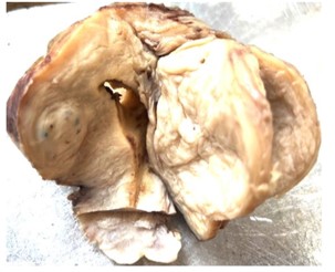
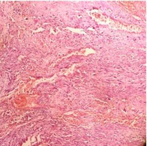
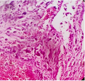
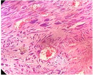
Leiomyoma with bizarre nuclei (LBN) is an uncommon variant of uterine smooth muscle neoplasm. A 52 year female presented with complained of intermittent pelvic pain, abnormal uterine bleeding, and abdominal discomfort of 6 month duration. On ultrasonic evaluation showed well defined uterine masses suggestive of leiomyomas. The systemic examinations were normal. On histopathologycal finding reported as uterine leiomyoma with bizarre nuclei. Patient responded well to treatment. On follow up there was no evidence of recurrence. We are presenting this uncommon case of leiomyoma with bizarre nuclei for its clinical, radiological and histopathological findings. On histopathological findings this tumor should be properly differentiated form various uterine spindle cell tumors with nuclear atypia for better management of patients.
References
- 1. Wei JJ. Leiomyoma with nuclear atypia: rare diseases that present a common diagnostic problem. Semin Diagn Pathol. 2022;39(3):187–200.
- 2. Kurman R, Carcangiu M, Herrington C, Young R. WHO Classification of Tumours of Female Reproductive Organs. Lyon: IARC Publications; 2014.
- 3. Board WCoTE. Female Genital Tumours. International Agency for Research on Cancer, World Health Organization; 2020.
- 4. Oliva E. Practical issues in uterine pathology from banal to bewildering: the remarkable spectrum of smooth muscle neoplasia. Mod Pathol. 2016;29 (Suppl 1):S104–S120.
- 5. Dafle VD, Kokode DN, Jagtap SV, More SS. Uterine Leiomyoma: A Clinicopathological Study in Hysterectomy Specimen Indian Journal of Pathology: Research and Practice. 2019,8, 4:502-506.
- 6. Croce S, Young RH, Oliva E.Uterine leiomyomas with bizarre nuclei: a clinicopathologic study of 59 cases. Am J Surg Pathol.201438: 1330-1339.
- 7. Jagtap SV, Phalke A, Kshirsagar NS, Jagtap SS, Nasre N. Uterine Lipoleiomyoma: a rare variant of benign uterine neoplasm. Int J Reprod Contracept Obstet Gynecol 2019;8:2553-5.
- 8. Pandis N, Hei S, Willén H, Bardi G, et al. Histologic cytogenetic correlations in uterine leiomyomas. International Journal of Gynecological Cancer 19911 (4): 163-68.
- 9. Travaglino A, Raffone A, Santoro A, et al. Risk of recurrence in uterine leiomyoma with bizarre nuclei: a systematic review and meta-analysis. Geburtshilfe Frauenheilkd. 2021;81(11):1217–1223.
- 10. Jagtap SV, Aher V, Gadhiya S, Jagtap SS. Gestational Trophoblastic Disease - Clinicopathological Study at Tertiary Care Hospital. J Clin Diagn Res. 2017; 11(8):EC27-EC30.
- 11. Anwar R, Tjandraprawira KD, Irawan B. Concurrent gestational trophoblastic neoplasia and large uterine fibroid in a nullipara - Case report. Annals of Medicine and Surgery.2022;77:103659.
Data Sharing Statement
There are no additional data available. All raw data and code are available upon request.
Funding
This research received no funding.
Author Contributions
All authors contributed significantly to the work and approve its publication.
Ethics Declaration
This article does not involve any human or animal subjects, and therefore does not require ethics approval.
Acknowledgements
We would like to express our gratitude to the patients, their families, and all those who have contributed to this study.
Conflicts of Interest
No conflicts of interest in this work.
About this article

Cite this article
Sunil V. Jagtap, Swati S Jagtap, Pranjal Shah, et al./ Leiomyoma with Bizarre Nuclei: An Uncommon Variant of Uterine Smooth Muscle Neoplasm/Indian Journal of Pathology: Research and Practice 2023;12(3) 105–108.
Licence:
Attribution-Non-commercial 4.0 International (CC BY-NC 4.0)This license
enables reusers to distribute, remix, adapt, and build upon the material in any
medium or format for noncommercial purposes only, and only so long as
attribution is given to the creator.
| Received | Accepted | Published |
|---|---|---|
| March 23, 2023 | June 15, 2023 | September 30, 2023 |
DOI: https://doi.org/10.21088/ijprp.2278.148X.12323.3
Keywords
Leiomyoma with bizarre nucleiSmooth muscle tumorsUterine tumorsHistopathologySearch for Similar Articles
Similar Articles
- Clinico-Pathological and Immunohistochemical Study of Primary CNS Tumors in a Te...
- The Role of SGLT2 Inhibitors in Cardiovascular Protection Beyond Diabetes: A Co...
- Epidermal Inclusion Cyst Presenting as 4th Ventricle Space Occupying Lesion: A R...
- Silent Retroperitoneal Seminoma Arising from an Occult Undescended Testis: Diagn...
- Scapular Metastasis of Oral Squamous Cell Carcinoma: A Case Report of an Unusual...
Article Level Metrics
Last UpdatedSaturday 17 October 2026, 10:30:26 (IST)
4177
Accesses
12
671
00
Citations
NA
NA
NA
Download citation
Article Keywords
Keyword Highlighting
Highlight selected keywords in the article text.
Timeline
| Received | March 23, 2023 |
| Accepted | June 15, 2023 |
| Published | September 30, 2023 |
licence
Attribution-Non-commercial 4.0 International (CC BY-NC 4.0)
This license
enables reusers to distribute, remix, adapt, and build upon the material in any
medium or format for noncommercial purposes only, and only so long as
attribution is given to the creator.