Full Text (PDF)
Indian Journal of Pathology: Research and Practice 15(2):p 101-110, May - Aug 2026. | DOI: https://doi.org/10.21088/ijprp.2278.148X.15226.1
Original Article
Clinico-Pathological and Immunohistochemical Study of Primary CNS Tumors in a Tertiary Care Centre
Shagufta Qadri, Kausar Tasnim, Maheshwari Veena, Sharma M. Raman
Author Information
Licence:
Attribution-Non-commercial 4.0 International (CC BY-NC 4.0)This license enables reusers to distribute, remix, adapt, and build upon the material in any medium or format for noncommercial purposes only, and only so long as attribution is given to the creator.
Indian Journal of Pathology: Research and Practice 15(2):p 101-110, May - Aug 2026. | DOI: https://doi.org/10.21088/ijprp.2278.148X.15226.1
How Cite This Article:
Kausar Tasnim, Shagufta Qadri, Maheshwari Veena. Clinico-Pathological and Immunohistochemical Study of Primary CNS Tumors in a Tertiary Care Centre. Ind Jr of Path: Res and Practice 2026; 15(2) 101-110.Timeline
Received : December 20, 2025
Accepted : January 25, 2026
Published : August 30, 2026
Abstract
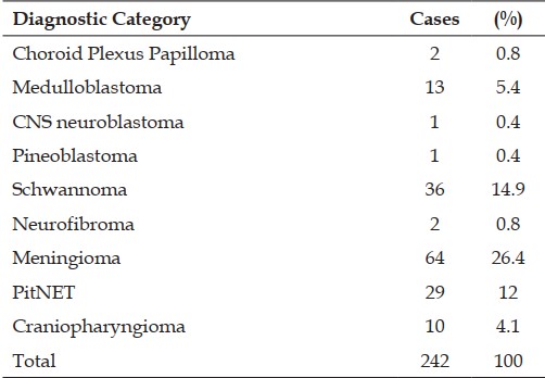
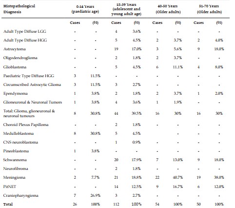
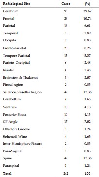
Background: Primary CNS tumors show wide clinical, histomorphological, and immunohistochemical variation. Distinguishing these lesions from each other and from reactive process can be challenging due to overlapping cell morphology. So, the diagnosis and categorization of primary CNS tumors, is essential for planning the management and to determine the prognosis. Objectives: To evaluate the clinico-radiological features in diagnosing CNS tumors; to study the occurrence of CNS tumors in relation to age, sex, and anatomical sites, and the histopathological features of various CNS tumors and to evaluate the role of immunohistochemistry in diagnosing as well as classifying various CNS tumors according to CNS5. Materials and Methods: This study included a total of 242 case’ biopsy of primary CNS tumors received in the Department of Pathology over 5 years from 2020 to 2024. All specimens were examined grossly, processed routinely, stained with H&E, and evaluated using a panel of IHC markers (IDH1, ATRX, p53, Ki-67, GFAP, Synaptophysin, EMA, PR, etc.). Results: Out of 242, the most common age group was 15-39 years with male: female ratio of 1.3:1 with most common site- cerebrum. Majority of the cases were non-malignant (61%), while 39% cases were malignant. The most frequent tumors were gliomas, followed by meningiomas, schwannoma, pituitary adenomas, and medulloblastomas. IHC significantly assisted in differentiating astrocytic tumors, subtyping meningiomas, and confirming embryonal tumors. Ki‑67 index correlated with the tumor grade. Conclusion: A combined clinico-pathological and IHC approach markedly improves diagnostic accuracy of primary CNS tumors.
References
- 1. Kanthikar S.N., Nikumbh D.B. and Dravid NV. Histopathological overview of central nervous system tumors in North Maharastra, India: A single center study. Ind J. Pathol and Oncol. 2017; 4(1): 80-4. DOI:10.18231/2394- 6792.2017.0016
- 2. Sung H., Ferlay J., Siegel R.L., Laversanne M., Soerjomataram I., Jemal A., et al. Global Cancer Statistics 2020: GLOBOCAN estimates of incidence and mortality worldwide for 36 cancers in 185 countries. CA Cancer J Clin. 2021; 71: 209-49. https://doi.org/10.3322/ caac.21660
- 3. Mondal S., Pradhan R., Pal S., Biswas B., Banerjee A., Bhattacharyya D. Clinicopathological pattern of brain tumors: A 3year study in a tertiary care hospital in India. Clin Cancer Investig J 2016; 5: 43740 DOI: 10.4103/22780513.197861
- 4. Naik S., Sahoo N., Mohanty B., Lenka A., Mohapatra A.S., Das P., et al. Histopathological spectrum of central nervous system lesions in a tertiary care hospital in Eastern India. J Evid Based Med Healthc 2021; 8(18): 1304-10. DOI: 10.18410/jebmh/2021/249
- 5. Ostrom Q.T., Francis S.S., Barnholtz-Sloan J.S. Epidemiology of brain and other CNS tumors. Curr Neurol Neurosci Rep. 2021; 21(12): 68. DOI: 10.1007/s11910-021-01152-9
- 6. Jaiswal J., Shastry A.H., Ramesh A., Chickabasaviah Y.T., Arimappamagan A., Santosh V. Spectrum of primary intracranial tumors at a tertiary care neurological institute: A hospital-based brain tumor registry. Neurol India 2016; 64: 494-50. DOI:10.4103/0028- 3886.181535
- 7. Louis D.N., Perry A., Wesseling P., Brat D.J., Cree I.A., Figarella-Branger D. et al. The 2021 WHO Classification of Tumors of the Central Nervous System: a summary. Neuro Oncol 2021; 23(8): 1231-51. DOI: 10.1093/neuonc/ noab106
- 8. Meel M., Choudhary N., Kumar M., Mathur K. Epidemiological Profiling and Trends of Primary Intracranial Tumors: A HospitalBased Brain Tumor Registry from a Tertiary Care Center. J Neurosci Rural Pract. 2021; 12(1): 96–101. DOI: 10.1055/s-0040-1721622
- 9. Price M., Ballard C., Benedetti J., Neff C., Cioffi G., Waite K.A., Kruchko C., Barnholtz-Sloan J.S., Ostrom Q.T. CBTRUS Statistical Report: Primary brain and other central nervous system tumors diagnosed in the United States in 2017– 2021. Neuro Oncol. 2024 Oct 6; 26 (Suppl 6): vi1– vi85. doi.org/10.1093/neuonc/noae145
- 10. Qadri S., Hasan M., Akhtar K. Magnitude of Neurogenic Tumor Burden in Pediatric Population: A Tertiary Care Center Study. J Pediatr Neurosci. 2017 Jul-Sep; 12(3): 222-226. DOI:10.4103/jpn.JPN_158_16
- 11. Thambi R., Kandamuthan S., Sainulabdeen S., Vilasiniamma L., Abraham T.R., Balakrishnan P.K. Histopathological Analysis of Brain Tumours-A Seven Year Study from a Tertiary Care Centre in South India. J Clin Diagn Res. 2017 Jun; 11(6): EC05-EC08. DOI: 10.7860/ JCDR/2017/25623.9990
- 12. Ozawa M., Brennan P.M., Zienius K., et al. The usefulness of symptoms alone or combined for general practitioners in considering the diagnosis of a brain tumour: a case-control study using the clinical practice research database (CPRD) (2000-2014). BMJ Open 2019; 9: e029686. DOI: 10.1136/bmjopen-2019-029686
- 13. Basu T., Das M., Debnath R., Bala A., Chatterjee U., Ghosh S. Clinicopathological profile of ependymomas with special reference to survival data - Experiences of a tertiary care center’. Indian J Pathol Microbiol. 2025 Jan 1; 68(1): 118-124. DOI: 10.4103/ijpm.ijpm_814_23
- 14. Anghileri E., Gaviani P., Amato A., Pollo B., Paterra R., Marchetti M., et al. Choroid plexus tumors in adults: a retrospective mono-institutional study. Neurol Sci. 2025 Apr; 46(4): 1859-1866. DOI: 10.1007/s10072-024-07894-x
- 15. Kool M., Korshunov A., Remke M., et al. Molecular subgroups of medulloblastoma: an international meta-analysis of transcriptome, genetic aberrations, and clinical data of WNT, SHH, Group 3, and Group 4 medulloblastomas. Acta Neuropathol. 2012 Apr; 123(4): 473-84 DOI: 10.1007/s00401-012-0958-8
- 16. Bianchi F., Tamburrini G., Gessi M., Frassanito P., Massimi L., Caldarelli M. Central nervous system (CNS) neuroblastoma. A case-based update. Childs Nerv Syst. 2018 May; 34(5): 817–823. DOI: 10.1007/s00381-018-3764-3
- 17. Jaju A., Hwang E.I., Kool M., Capper D., Chavez L., Brabetz S. MRI Features of Histologically Diagnosed Supratentorial Primitive Neuroectodermal Tumors and Pineoblastomas in Correlation with Molecular Diagnoses and Outcomes: A Report from the Children’s Oncology Group ACNS0332 Trial. AJNR Am J Neuroradiol. 2019 Nov; 40(11): 1796-1803. DOI: 10.3174/ajnr.A6253
- 18. Saeger W., Lüdecke D.K., Buchfelder M., Fahlbusch R., Quabbe H.J., Petersenn S. Pathohistological classification of pituitary tumors: 10 years of experience with the German Pituitary Tumor Registry. Eur J Endocrinol. 2007 Feb; 156(2): 203–216. DOI: 10.1530/eje.1.02326
Data Sharing Statement
There are no additional data available. All raw data and code are available upon request.
Funding
This research received no funding.
Author Contributions
All authors contributed significantly to the work and approve its publication.
Ethics Declaration
This article does not involve any human or animal subjects, and therefore does not require ethics approval.
Acknowledgements
We would like to express our gratitude to the patients, their families, and all those who have contributed to this study.
Conflicts of Interest
No conflicts of interest in this work.
About this article

Cite this article
Kausar Tasnim, Shagufta Qadri, Maheshwari Veena. Clinico-Pathological and Immunohistochemical Study of Primary CNS Tumors in a Tertiary Care Centre. Ind Jr of Path: Res and Practice 2026; 15(2) 101-110.
Licence:
Attribution-Non-commercial 4.0 International (CC BY-NC 4.0)This license enables reusers to distribute, remix, adapt, and build upon the material in any medium or format for noncommercial purposes only, and only so long as attribution is given to the creator.
| Received | Accepted | Published |
|---|---|---|
| December 20, 2025 | January 25, 2026 | August 30, 2026 |
DOI: https://doi.org/10.21088/ijprp.2278.148X.15226.1
Keywords
GliomasImmunohistochemistryMedulloblastomasMeningiomasPituitary adenomasSchwannomaSearch for Similar Articles
Similar Articles
- The Role of SGLT2 Inhibitors in Cardiovascular Protection Beyond Diabetes: A Co...
- Epidermal Inclusion Cyst Presenting as 4th Ventricle Space Occupying Lesion: A R...
- Silent Retroperitoneal Seminoma Arising from an Occult Undescended Testis: Diagn...
- Scapular Metastasis of Oral Squamous Cell Carcinoma: A Case Report of an Unusual...
- Adrenocortical Carcinoma Presenting as a Retroperitoneal Mass: Diagnostic Challe...
Article Level Metrics
Last UpdatedSunday 18 October 2026, 14:54:49 (IST)
4203
Accesses
32
671
00
Citations
NA
NA
NA
Download citation
Article Keywords
Keyword Highlighting
Highlight selected keywords in the article text.
Timeline
| Received | December 20, 2025 |
| Accepted | January 25, 2026 |
| Published | August 30, 2026 |
licence
Attribution-Non-commercial 4.0 International (CC BY-NC 4.0)
This license enables reusers to distribute, remix, adapt, and build upon the material in any medium or format for noncommercial purposes only, and only so long as attribution is given to the creator.