Heading
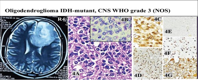
Description: Fig. R4 MRI showed an ill-defined right frontal intra-axial mass with T2/FLAIR hyperintensity, cystic areas, vasogenic edema, and mild midline shift; histomorphology (Fig. 4A&B, H&E,10X) showed diffuse round cells with perinuclear halos and brisk mitoses (40X). IHC showed IDH-mutation (Fig. 4C, 40X), ATRX-retained (Fig. 4D, 40X), p53-negative (Fig. 4E, 10X), Ki-67 (30–40%, 10X) (Fig. 4F, 10X), and GFAP positivity (Fig. 4G, 40X) in tumor cells