Full Text (PDF)
Indian Journal of Forensic Medicine and Pathology 13(1):p 74-82, January-March 2020. | DOI: https://doi.org/10.21088/ijfmp.0974.3383.13120.11
Original Article
Assessment of c-erbB2 Expression by IHC and FISH in Invasive Breast Cancer— A Comparative Study: Experience from a Single Institute
Swati Sharma, Nilay Nishith, Ranjini Kudva, Shankar M Bakkannavar
Author Information
Licence:
Attribution-Non-commercial 4.0 International (CC BY-NC 4.0)This license enables reusers to distribute, remix, adapt, and build upon the material in any medium or format for noncommercial purposes only, and only so long as attribution is given to the creator.
Indian Journal of Forensic Medicine and Pathology 13(1):p 74-82, January-March 2020. | DOI: https://doi.org/10.21088/ijfmp.0974.3383.13120.11
How Cite This Article:
Nishith N, Sharma S, Kudva R, et al. Assessment of c-erbB2 Expression by IHC and FISH in Invasive Breast Cancer— A Comparative Study: Experience from a Single Institute. Indian J Forensic Med Pathol. 2020;13(1):74–82.Timeline
Received : September 16, 2019
Accepted : November 28, 2019
Published : December 30, 2019
Abstract
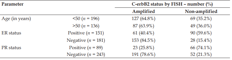
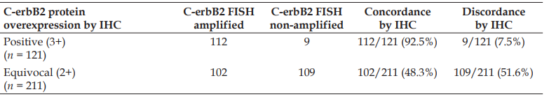
Introduction: An accurate assessment of c-erbB2 expression in invasive breast cancer (IBC) has become crucial to precisely recognize the candidates to be treated with Trastuzumab. Presently, fluorescence in situ hybridization (FISH) and immunohistochemistry (IHC) are most commonly employed methods for evaluating c-erbB2 status. Recent literature has documented a strong correlation between the two c-erbB2 diagnostic analyzes. However, discordance between both the assays has been rarely reported. Therefore, we aimed to compare and correlate FISH and IHC results for c-erbB2 expression in Indian breast cancer patients. Material and methods: A total of 388 formalin fixed, paraffin embedded blocks of invasive breast cancer were retrospectively evaluated for c-erbB2 status by IHC (DAKO) and FISH (PathVysion dual-probe system) and results were compared. Results: 92.5% cases with IHC 3+ score showed significant concordance with the FISH results; while c-erbB2 gene amplification was noted in 48.3% of IHC 2+ cases. A large number of referral cases in the study group and variation in pre-analytical and analytical factors have attributed in escalating the number of indeterminate cases expressing c-erbB2 gene amplification. Additionally, an inverse correlation was revealed between ER/ PR expression and c-erbB2 status. Conclusion: The results of the current study established a high degree of concordance between IHC and FISH in Indian breast cancer patients with 3+ immunoreactivity. However, reflex testing by FISH is recommended for IHC equivocal cases in order to avoid false results related to technical and interpretation errors, usually encountered while performing an immunohistochemical assessment.
References
- 1. Ménard S, Pupa SM, Campiglio M, et al. Biologic and therapeutic role of HER2 in cancer. Oncogene. 2003 Sep 29;22(42):6570–8.
- 2. Iqbal N, Iqbal N. Human Epidermal Growth Factor Receptor 2 (HER2) in Cancers: Overexpression and Therapeutic Implications. Mol Biol Int. 2014;2014:852748.
- 3. Gravalos C, Jimeno A. HER2 in gastric cancer: a new prognostic factor and a novel therapeutic target. Ann Oncol. 2008 Sep;19(9):1523–9.
- 4. Emde A, Köstler WJ, Yarden Y. Therapeutic strategies and mechanisms of tumorigenesis of HER2-overexpressing breast cancer. Crit Rev Oncol Hematol. 2012 Dec;84 Suppl 1:e49–57.
- 5. Mitri Z, Constantine T, O’Regan R. The HER2 Receptor in Breast Cancer: Pathophysiology, Clinical Use, and New Advances in Therapy. Chemother Res Pract. 2012;2012:743193.
- 6. Panjwani P, Epari S, Karpate A, et al. Assessment of HER-2/neu status in breast cancer using fluorescence in situ hybridization and immunohistochemistry: Experience of a tertiary cancer referral centre in India. Indian J Med Res. 2010 Sep;132:287–94.
- 7. Gutierrez C, Schiff R. HER2: biology, detection, and clinical implications. Arch Pathol Lab Med. 2011 Jan;135(1):55–62.
- 8. Diaz NM. Laboratory testing for HER2/neu in breast carcinoma: an evolving strategy to predict response to targeted therapy. Cancer Control. 2001 Sep-Oct;8(5):415–8.
- 9. Varga Z, Noske A, Ramach C, et al. Assessment of HER2 status in breast cancer: overall positivity rate and accuracy by fluorescence in situ hybridization and immunohistochemistry in a single institution over 12 years: a quality control study. BMC Cancer. 2013 Dec 30;13:615.
- 10. Masood S. Prognostic/predictive factors in breast cancer. Clin Lab Med. 2005 Dec;25(4):809–25.
- 11. Gunnarsson C, Jansson T, Holmlund B, Olsson H. Methods for evaluating HER2 status in breast cancer: comparison of IHC, FISH, and real-time PCR analysis of formalin-fixed paraffin-embedded tissue. Pathology and Laboratory Medicine International. 2013 Sept;5:31–37.
- 12. Nichols DW, Wolff DJ, Self S, et al. A testing algorithm for determination of HER2 status in patients with breast cancer. Ann Clin Lab Sci. 2002 Winter;32(1):3–11.
- 13. Payne SJ, Bowen RL, Jones JL, et al. Predictive markers in breast cancer-the present. Histopathology. 2008 Jan;52(1):82–90.
- 14. Wolff AC, Hammond ME, Schwartz JN, et al. American Society of Clinical Oncology/College of American Pathologists guideline recommendations for human epidermal growth factor receptor 2 testing in breast cancer. J Clin Oncol. 2007 Jan 1;25(1):118–45.
- 15. Yamauchi H, Stearns V, Hayes DF. When is a tumor marker ready for prime time? A case study of c-erbB-2 as a predictive factor in breast cancer. J Clin Oncol. 2001 Apr 15;19(8):2334–56.
- 16. Allred DC. Issues and updates: evaluating estrogen receptor-alpha, progesterone receptor, and HER2 in breast cancer. Mod Pathol. 2010 May;23 Suppl 2:S52–9.
- 17. Sauter G, Lee J, Bartlett JM, et al. Guidelines for human epidermal growth factor receptor 2 testing: biologic and methodologic considerations. J Clin Oncol. 2009 Mar 10;27(8):1323–33.
- 18. Higa GM, Fell RG. Sex hormone receptor repertoire in breast cancer. Int J Breast Cancer. 2013;2013:284036.
- 19. Wolff AC, Hammond MEH, Hicks DG, et al. Recommendations for human epidermal growth factor receptor 2 testing in breast cancer: American Society of Clinical Oncology/College of American Pathologists clinical practice guideline update. Arch Pathol Lab Med. 2014 Feb;138(2):241–56.
- 20. Wolff AC, Hammond MEH, Allison KH, et al. Human Epidermal Growth Factor Receptor 2 Testing in Breast Cancer: American Society of Clinical Oncology/College of American Pathologists Clinical Practice Guideline Focused Update. Arch Pathol Lab Med. 2018 Nov;142(11):1364–82.
- 21. Allred DC, Harvey JM, Berardo M, et al. Prognostic and predictive factors in breast cancer by immunohistochemical analysis. Mod Pathol. 1998 Feb;11(2):155–68.
- 22. Shokouh TZ, Ezatollah A, Barand P. Interrelationships Between Ki67, HER2/neu, p53, ER, and PR Status and Their Associations With Tumor Grade and Lymph Node Involvement in Breast Carcinoma Subtypes: Retrospective Observational Analytical Study. Medicine (Baltimore). 2015 Aug;94(32):e1359.
- 23. Payandeh M, Sadeghi M, Sadeghi E, et al. Is There any Concordance between of IHC with FISH in HER2-Positive Breast Cancer Patients? Int J Hematol Oncol Stem Cell Res. 2017 Jan 1;11(1):43–48.
- 24. Eswarachary V, Mohammed IG, Jayanna PK, et al. HER2/neu Testing In 432 Consecutive Breast Cancer Cases using FISH and IHC - A Comparative Study. J Clin Diagn Res. 2017 Apr;11(4):EC01-EC05.
- 25. Murthy SS, Sandhya DG, Ahmed F, et al. Assessment of HER2/Neu status by fluorescence in situ hybridization in immunohistochemistry-equivocal cases of invasive ductal carcinoma and aberrant signal patterns: a study at a tertiary cancer center. Indian J Pathol Microbiol. 2011 Jul-Sep;54(3):532–8.
- 26. Jimenez RE, Wallis T, Tabasczka P, Visscher DW. Determination of HER-2/Neu status in breast carcinoma: comparative analysis of immunohistochemistry and fluorescent in situ hybridization. Mod Pathol. 2000 Jan;13(1):37–45.
- 27. Mostafa NA, Eissa SS, Belal DM, Shoman SH. Assessment of HER-2/neu gene amplification status in breast carcinoma with equivocal 2+ Her2/neu immunostaining. J Egypt Natl Canc Inst. 2011 Mar;23(1):41–6.
- 28. Crowe JP, Patrick RJ, Rybicki LA, et al. A data model to predict HER2 status in breast cancer based on the clinical and pathologic profiles of a large patient population at a single institution. Breast. 2006 Dec;15(6):728–35.
- 29. Prati R, Apple SK, He J, et al. Histopathologic characteristics predicting HER-2/neu amplification in breast cancer. Breast J. 2005 Nov-Dec;11(6):433–9.
- 30. Massarweh S, Schiff R. Resistance to endocrine therapy in breast cancer: exploiting estrogen receptor/growth factor signaling crosstalk. Endocr Relat Cancer. 2006 Dec;13 Suppl 1:S15–24.
- 31. Shou J, Massarweh S, Osborne CK, et al. Mechanisms of tamoxifen resistance: increased estrogen receptor-HER2/neu cross-talk in ER/HER2-positive breast cancer. J Natl Cancer Inst. 2004 Jun 16;96(12):926–35.
- 32. Osborne CK, Bardou V, Hopp TA, et al. Role of the estrogen receptor coactivator AIB1 (SRC-3) and HER-2/neu in tamoxifen resistance in breast cancer. J Natl Cancer Inst. 2003 Mar 5;95(5):353–61.
- 33. Rakha EA, Reis-Filho JS, Ellis IO. Basal-like breast cancer: a critical review. J Clin Oncol. 2008 May 20;26(15):2568–81.
- 34. Jacobs TW, Gown AM, Yaziji H, et al. Comparison of fluorescence in situ hybridization and immunohistochemistry for the evaluation of HER-2/neu in breast cancer. J Clin Oncol. 1999 Jul;17(7):1974–82.
- 35. Owens MA, Horten BC, Da Silva MM. HER2 amplification ratios by fluorescence in situ hybridization and correlation with immunohistochemistry in a cohort of 6556 breast cancer tissues. Clin Breast Cancer. 2004 Apr;5(1):63–9.
- 36. Birner P, Oberhuber G, Stani J, et al. Evaluation of the United States Food and Drug Administration-approved scoring and test system of HER-2 protein expression in breast cancer. Clin Cancer Res. 2001 Jun;7(6):1669–75.
- 37. Makroo RN, Chowdhry M, Kumar M, et al. Correlation between HER2 gene amplification and protein overexpression through fluorescence in situ hybridization and immunohistochemistry in breast carcinoma patients. Indian J Pathol Microbiol. 2012 Oct-Dec;55(4):481–4.
- 38. Wang L, Wang X, Nie X, et al. Comparison of fluorescence in situ hybridization and immunohistochemistry for assessment of HER-2 status in breast cancer patients. J Huazhong Univ Sci Technolog Med Sci. 2009 Jun;29(3):354–8.
- 39. Hammock L, Lewis M, Phillips C, et al. Strong HER-2/neu protein overexpression by immunohistochemistry often does not predict oncogene amplification by fluorescence in situ hybridization. Hum Pathol. 2003 Oct;34(10):1043–7.
- 40. Perez EA, Suman VJ, Davidson NE, et al. HER2 testing by local, central, and reference laboratories in specimens from the North Central Cancer Treatment Group N9831 intergroup adjuvant trial. J Clin Oncol. 2006 Jul 1;24(19):3032–8.
- 41. Tsuda H, Akiyama F, Terasaki H, et al. Detection of HER-2/neu (c-erb B-2) DNA amplification in primary breast carcinoma. Interobserver reproducibility and correlation with immunohistochemical HER-2 overexpression. Cancer. 2001 Dec 15;92(12):2965–74.
- 42. Tubbs RR, Pettay JD, Roche PC, et al. Discrepancies in clinical laboratory testing of eligibility for trastuzumab therapy: apparent immunohistochemical false-positives do not get the message. J Clin Oncol. 2001 May 15;19(10):2714–21.
- 43. Portier BP, Wang Z, Downs-Kelly E, et al. Delay to formalin fixation ‘cold ischemia time’: effect on ERBB2 detection by in-situ hybridization and immunohistochemistry. Mod Pathol. 2013 Jan;26(1):1–9.
- 44. Lewis JT, Ketterling RP, Halling KC, et al. Analysis of intratumoral heterogeneity and amplification status in breast carcinomas with equivocal (2+) HER-2 immunostaining. Am J Clin Pathol. 2005 Aug;124(2):273–81.
- 45. Yaziji H, Goldstein LC, Barry TS, et al. HER-2 testing in breast cancer using parallel tissue-based methods. JAMA. 2004 Apr 28;291(16):1972–7.
- 46. Ward CJ, Green VL. Risk Management and Medico-Legal Issues in Breast Cancer. Clin Obstet Gynecol. 2016 Jun;59(2):439–46..
Data Sharing Statement
There are no additional data available. All raw data and code are available upon request.
Funding
This research received no funding.
Author Contributions
Whether all authors contributed significantly to the work and approve its publication.
Ethics Declaration
This article does not involve any human or animal subjects, and therefore does not require ethics approval.
Acknowledgements
We would like to express our gratitude to the patients, their families, and all those who have contributed to this study.
Conflicts of Interest
The authors report no conflicts of interest in this work.
About this article
Cite this article
Nishith N, Sharma S, Kudva R, et al. Assessment of c-erbB2 Expression by IHC and FISH in Invasive Breast Cancer— A Comparative Study: Experience from a Single Institute. Indian J Forensic Med Pathol. 2020;13(1):74–82.
Licence:
Attribution-Non-commercial 4.0 International (CC BY-NC 4.0)This license enables reusers to distribute, remix, adapt, and build upon the material in any medium or format for noncommercial purposes only, and only so long as attribution is given to the creator.
| Received | Accepted | Published |
|---|---|---|
| September 16, 2019 | November 28, 2019 | December 30, 2019 |
DOI: https://doi.org/10.21088/ijfmp.0974.3383.13120.11
Keywords
Breast cancerc-erbB2ConcordanceFluorescent in situ hybridization(FISH)Immunohistochemistry (IHC)Search for Similar Articles
Similar Articles
- Analysis of Death Due to Pulmonary Embolism, a Case Series
- Suspected Adverse Event Following Immunization with Multisystem Inflammatory Res...
- Drone: A Smart Intelligent Framework Aiding Forensic Investigations
- Assessment of Knowledge on Sexual Assault Forensic Examination among Nurses in I...
- An Analysis of the Medico-Legal Aspects and Trends in Sexual Offense Cases at a...
Article Level Metrics
Last UpdatedThursday 09 July 2026, 08:57:02 (IST)
7829
Accesses
6
2153
00
Citations
NA
NA
NA
Download citation
Article Keywords
Keyword Highlighting
Highlight selected keywords in the article text.
Timeline
| Received | September 16, 2019 |
| Accepted | November 28, 2019 |
| Published | December 30, 2019 |
licence
Attribution-Non-commercial 4.0 International (CC BY-NC 4.0)
This license enables reusers to distribute, remix, adapt, and build upon the material in any medium or format for noncommercial purposes only, and only so long as attribution is given to the creator.