Full Text (PDF)
Indian Journal of Forensic Medicine and Pathology 16(4):p 253-259, October-December 2023. | DOI: https://doi.org/10.21088/ijfmp.0974.3383.16423.4
Original Article
A Orthopantomography Study of Age Estimation Based on PeriodontalLigament Visibility of MandibularThird Molars in Chennai Population: An Aid in Forensics
Author Information
Licence:
Attribution-Non-commercial 4.0 International (CC BY-NC 4.0)This license enables reusers to distribute, remix, adapt, and build upon the material in any medium or format for noncommercial purposes only, and only so long as attribution is given to the creator
Indian Journal of Forensic Medicine and Pathology 16(4):p 253-259, October-December 2023. | DOI: https://doi.org/10.21088/ijfmp.0974.3383.16423.4
How Cite This Article:
Rangeela M, Dharman S. A orthopantomography study of age estimation based on periodontal ligament visibility of mandibular third molars in Chennai population: an aid in forensics. Indian J Forensic Med Pathol. 2023;16(4):253-259.Timeline
Received : September 04, 2023
Accepted : December 29, 2023
Published : December 31, 2023
Abstract
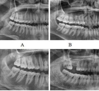
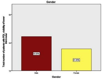
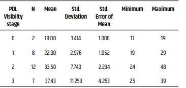
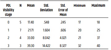
Forensic age determination has evolved to be intriguingly signifi cant and relates to the challenges originating from globalisation as a result of a growing amount of foreign nationals with questionable birth date information and to identify victims who are unknown. Aim of this research was to evaluate the estimation of age through visualisation of periodontal ligament (PDL) around fully mineralised lower third molars in the Chennai population, and also to assess the 18-year age threshold using PDL visibility. The research was a retrospective analysis that involved 100 OPGs (orthopantomographs) from the archives of the Department of Oral and Maxillofacial Radiology from 2021 to 2022 were analysed to determine PDL present around the mandibular third molars. Grading each mandibular third molar was done by two observers. IBM SPSS version 23 was used to statistically evaluate the gathered data. Among 100 study subjects, the mean age group was found to be 24.2 years. The frequency distribution of age was inferred from that of the study participants, 15% were under the age of 18, and 85% were above the age of 18. Males and girls with both lower third molars on the left and right had grades of stage 1 (34%), stage 2 (31%), and stage 3 (20%) at the age of 18. The 18-year threshold can be established by using periodontal ligament visibility, according to our fi ndings, starting from Stage 1. The relationship between the variables was examined using the Chi square test, which produced a very signifi cant p value of 0.00. Radiographic age evaluation was a simple, repeatable, non-invasive technique that can be applied to both living and deceased people. Our research indicated that less than 18 years old was the minimal age for Stage 0 in both males and females. At the onset of stages 1, 2, and 3, all males and females were at least eighteen years old. Stages 1, 2, or 3 indicate that the person is older than 18. The populace of Chennai would benefi t from this innovative method of age estimation for use in forensics.
References
- 1. Schmeling A, Grundmann C, Fuhrmann A, Kaatsch HJ, Knell B, Ramsthaler F, Reisinger W, Riepert T, Ritz-Timme S, Rösing FW, Rötzscher K. Criteria for age estimation in living individuals. International journal of legal medicine. 2008 Nov;122:457-60.
- 2. Yang Z, Geng K, Liu Y, Sun S, Wen D, Xiao J, Zheng Y, Cai J, Zha L, Liu Y. Accuracy of the Demirjian and Willems methods of dental age estimation for children from central southern China. International Journal of Legal Medicine. 2019 Mar 5;133:593-601.
- 3. Akshaya A, Arthanari A. Dental Age Estimation of Lower Left Third Molar (38) Using Modified Willems Method in South Indian Population. Journal of Research in Medical and Dental Science. 2021 Oct;9(10):77-82.
- 4. Caldas IM, Júlio P, Simões RJ, Matos E, Afonso A, Magalhães T. Chronological age estimation based on third molar development in a Portuguese population. International journal of legal medicine. 2011 Mar;125:235-43.5.
- 5. Pérez-Mongiovi D, Teixeira A, Caldas IM. The radiographic visibility of the root pulp of the third lower molar as an age marker. Forensic science, medicine, and pathology. 2015 Sep;11:339-44.
- 6. Garamendi PM, Landa MI, Botella MC, Alemán I. Forensic age estimation on digital x-ray images: medial epiphyses of the clavicle and rst rib ossication in relation to chronological age. J Forensic Sci. 2011;56:S3-S12.
- 7. Alex A, Mohanraj KG. Morphological and morphometrical analysis of typical cervical vertebrae and their clinical implications. Drug Invention Today. 2019 Sep 15;12(9).
- 8. Dharman S. Assessment of Mandibular Condylar Morphology Using Digital Orthopantomogram in Chennai Population. Indian Journal of Forensic Medicine & Toxicology. 2020 Jul 1;14(3).
- 9. Demirjian A, Goldstein H, Tanner JM (1973) A new system of dental age.
- 10. Ravikumar H, Arthanari A. Dental Age Estimation of Upper Left Third Molar (28) Using Willems Method. Journal of Research in Medical and Dental Science. 2022 Jan;10(1):366-70.
- 11. Kvaal SI, Kolltveit KM, Thomsen IO, Solheim T. Age estimation of adults from dental radiographs. Forensic science international. 1995 Jul 28;74(3):175-85.
- 12. Olze A, Pynn BR, Kraul V, Schulz R, Heinecke A, Pfeiffer H, Schmeling A. Studies on the chronology of third molar mineralization in First Nations people of Canada. International journal of legal medicine. 2010 Sep;124:433-7.
- 13. Timme M, Timme WH, Olze A, Ottow C, Ribbecke S, Pfeiffer H, et al. The chronology of the radiographic visibility of the periodontal ligament and the root pulp in the lower third molars. Sci Justice 2017;57(4):257–61.
- 14. Guo YC, Wang YH, Olze A, Schmidt S, Schulz R, Pfeiffer H, Chen T, Schmeling A. Dental age estimation based on the radiographic visibility of the periodontal ligament in the lower third molars: application of a new stage classification. International Journal of Legal Medicine. 2020 Jan;134:369-74.
- 15. Sequeira CD, Teixeira A, Caldas IM, Afonso A, Pérez-Mongiovi D. Age estimation using the radiographic visibility of the periodontal ligament in lower third molars in a Portuguese population. Journal of clinical and experimental dentistry. 2014 Dec; 6(5):e546.
- 16. Vora S, Kardjokar F, Sansare K, Patankar S. Age estimation based on radiographic visibility of periodontal ligament surrounding mandibular third molars-A retrospective study. J Forensic Sci & Criminal Inves. 2019;11(5):1-4.
- 17. Nandita R, Arthanari A. OPG and CBCT-A More Reliable Source for Human Identification? Perception Based Web Survey among Dentist. Journal of Research in Medical and Dental Science. 2022 Jan;10(1):120-8.
- 18. Bosmans N, Ann P, Aly M, Willems G. The application of Kvaal’s dental age calculation technique on panoramic dental radiographs. Forensic SciInt 2005;153(2-3):208–12.
- 19. Agarwal N, Ahuja P, Sinha A, Singh A. Age estimation using maxillary central incisors: A radiographic study. J Forensic Dent Sci 2012;4(2):97–100.
- 20. Sequeira CD, Teixeira A, Caldas IM, Afonso A, Pérez-Mongiovi D. Age estimation using the radiographic visibility of the periodontal ligament in lower third molars in a Portuguese population. Journal of clinical and experimental dentistry. 2014 Dec; 6(5):e546.
- 21. Demirjian A, Goldstein H, Tanner JM. A new system of dental age assessment. Hum Biol 1973;45(2):211–27.
- 22. Karaarslan B, Karaarslan ES, Ozsevik AS, Ertas E. Age estimation for dental patients using orthopantomographs. European journal of dentistry. 2010 Oct;4(04):389-94.
- 23. Garamendi P, Landa MI, Ballesteros J, Solano MA. Reliability of the methods applied to assess age minority in living subjects around 18 years old: a survey on a Moroccan origin population. Forensic science international. 2005 Nov 10;154(1):3-12.
- 24. Orhan K, Ozer L, Orhan AI, Dogan S, Paksoy CS. Radiographic evaluation of third molar development in relation to chronological age among Turkish children and youth. Forensic science international. 2007 Jan 5;165(1):46-51
Data Sharing Statement
There are no additional data available. All raw data and code are available upon request.
Funding
This research received no funding.
Author Contributions
All authors contributed significantly to the work and approve its publication.
Ethics Declaration
This article does not involve any human or animal subjects, and therefore does not require ethics approval.
Acknowledgements
We would like to express our gratitude to the patients, their families, and all those who have contributed to this study.
Conflicts of Interest
No conflicts of interest in this work.
About this article

Cite this article
Rangeela M, Dharman S. A orthopantomography study of age estimation based on periodontal ligament visibility of mandibular third molars in Chennai population: an aid in forensics. Indian J Forensic Med Pathol. 2023;16(4):253-259.
Licence:
Attribution-Non-commercial 4.0 International (CC BY-NC 4.0)This license enables reusers to distribute, remix, adapt, and build upon the material in any medium or format for noncommercial purposes only, and only so long as attribution is given to the creator
| Received | Accepted | Published |
|---|---|---|
| September 04, 2023 | December 29, 2023 | December 31, 2023 |
DOI: https://doi.org/10.21088/ijfmp.0974.3383.16423.4
Keywords
Periodontal ligamentForensicMandibular third molarSearch for Similar Articles
Similar Articles
- Diatom Analysis in Thermophilic water bodies: A Study on Hot Water Springs of Ma...
- Morphometric Study of Supra-meatal Triangle and Mastoid process in the Adult dry...
- Histopathological Spectrum of Autopsy Cases: A One Year Retrospective Observatio...
- Forensic Differentiation of Manner of Death in Railway Fatalities: A FourCase Se...
- Understanding assault Patterns: A Cross-sectional study of Cases at Chri, Kelamb...
Article Level Metrics
Last UpdatedMonday 19 October 2026, 10:57:09 (IST)
16327
Accesses
44
3215
00
Citations
NA
NA
NA
Download citation
Article Keywords
Keyword Highlighting
Highlight selected keywords in the article text.
Timeline
| Received | September 04, 2023 |
| Accepted | December 29, 2023 |
| Published | December 31, 2023 |
licence
Attribution-Non-commercial 4.0 International (CC BY-NC 4.0)
This license enables reusers to distribute, remix, adapt, and build upon the material in any medium or format for noncommercial purposes only, and only so long as attribution is given to the creator