Full Text (PDF)
Indian Journal of Obstetrics and Gynecology 10(2):p 85-90, April – June 2022. | DOI: https://doi.org/10.21088/ijog.2321.1636.10222.9
Original Article
Uterine Artery Doppler At 11-14 weeks in Prediction of Preeclampsia
K Padma, G Soruba Rani, Nancy F
Author Information
Licence:
Attribution-Non-commercial 4.0 International (CC BY-NC 4.0)This license
enables reusers to distribute, remix, adapt, and build upon the material in any
medium or format for noncommercial purposes only, and only so long as
attribution is given to the creator.
Indian Journal of Obstetrics and Gynecology 10(2):p 85-90, April – June 2022. | DOI: https://doi.org/10.21088/ijog.2321.1636.10222.9
How Cite This Article:
G Soruba Rani, K Padma, Nancy F/Uterine Artery Doppler At 11-14 weeks in Prediction of Preeclampsia/Indian J Obstet Gynecol. 2022;10(2):85–90.Timeline
Received : December 03, 2021
Accepted : December 04, 2022
Published : June 30, 2022
Abstract
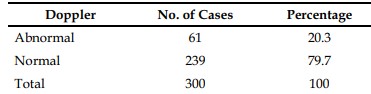
Identification of women at high risk for preeclampsia improves pregnancy outcome, early prediction of preeclampsia prevents dreadful maternal complication thereby reduces maternal mortality & morbidity and fetal complication. A prospective study conducted in Govt. Rajaji Hospital Madurai from January 2021 – September 2021. 300 antenatal women were interrogated between 11-14 weeks of GA with uterine artery Doppler. Out of which 55.3% of High risk women were predicted as preeclampsia & 27% of low risk women were predicted as preeclampsia. These patients were treated with T.Aspirin. So that maternal & perinatal morbidity & mortality reduced.
References
- 1. Uterine artery Doppler velocimetry in relation to trophoblast migration into the myometrium of the placental bed. Obstet Gynecol. 1995;85
- 2. Schulman h, Fleischer A, Farmakides G, Bracero L, Grunfeld L, Development of uterine artery compliance in pregnancy as detected by Doppler ultrasound. Am J Obstet Gynecol.1986;155:1031-6.
- 3. Gomez O, Maritinez JM, Fifueras F, et al. Uterine artery Doppler at 11-14 weeks of gestation toscreen for hypertensive disorders and associated complications in an unselected population. Ultrasound Obstet Gynecol. 2005; 26:490-4.
- 4. Papageorghiou AT, Yu CK, Erasmus IE, Cuckle HS, Nicolaides KH. Assessment of risk for the development of preeclampsia by maternal characteristics and uterine artery Doppler. BJOG.2005;112:703-9.
- 5. Cnossen J, Morris R, Riet G, et al. Use of uterine artery Doppler ultrasonography to predict preeclampsia and intrauterine growth restriction: a systematic review and bivariable meta-analysis, CMAJ.2008;178:701-11
Data Sharing Statement
There are no additional data available. All raw data and code are available upon request.
Funding
This research received no funding.
Author Contributions
All authors contributed significantly to the work and approve its publication.
Ethics Declaration
This article does not involve any human or animal subjects, and therefore does not require ethics approval.
Acknowledgements
We would like to express our gratitude to the patients, their families, and all those who have contributed to this study.
Conflicts of Interest
No conflicts of interest in this work.
About this article

Cite this article
G Soruba Rani, K Padma, Nancy F/Uterine Artery Doppler At 11-14 weeks in Prediction of Preeclampsia/Indian J Obstet Gynecol. 2022;10(2):85–90.
Licence:
Attribution-Non-commercial 4.0 International (CC BY-NC 4.0)This license
enables reusers to distribute, remix, adapt, and build upon the material in any
medium or format for noncommercial purposes only, and only so long as
attribution is given to the creator.
| Received | Accepted | Published |
|---|---|---|
| December 03, 2021 | December 04, 2022 | June 30, 2022 |
DOI: https://doi.org/10.21088/ijog.2321.1636.10222.9
Keywords
PreeclampsiaIUGRUterine Artery DopplerSearch for Similar Articles
Similar Articles
- Fibromyoma: A Clinical Dilemma
- Optimizing Obstetric Care: A Review of Indications and Outcomes for First-Time C...
- A Rare Case of Geriatric Endometrial Adenocarcinoma Presenting with Nonpuerperal...
- Relationship between Hypothyroidism and Gestational Diabetes Mellitus: A Prospec...
- Influence of Maternal Anthropometry and Hemoglobin on Neonatal Anthropometry
Article Level Metrics
Last UpdatedTuesday 08 September 2026, 16:16:35 (IST)
3505
Accesses
7
580
00
Citations
NA
NA
NA
Download citation
Article Keywords
Keyword Highlighting
Highlight selected keywords in the article text.
Timeline
| Received | December 03, 2021 |
| Accepted | December 04, 2022 |
| Published | June 30, 2022 |
licence
Attribution-Non-commercial 4.0 International (CC BY-NC 4.0)
This license
enables reusers to distribute, remix, adapt, and build upon the material in any
medium or format for noncommercial purposes only, and only so long as
attribution is given to the creator.