Full Text (PDF)
Indian Journal of Pathology: Research and Practice 12(4):p 143-146, October-December 2023. | DOI: https://doi.org/10.21088/ijprp.2278.148X.12423.4
Case Report
Primary Neuroendocrine Tumour of Extrahepatic Bile Duct: A Rarity
Neha Sharma, Asha Mary George, Anshika Rai
Author Information
Licence:
Attribution-Non-commercial 4.0 International (CC BY-NC 4.0)This license
enables reusers to distribute, remix, adapt, and build upon the material in any
medium or format for noncommercial purposes only, and only so long as
attribution is given to the creator.
Indian Journal of Pathology: Research and Practice 12(4):p 143-146, October-December 2023. | DOI: https://doi.org/10.21088/ijprp.2278.148X.12423.4
How Cite This Article:
Asha Mary George, Anshika Rai, Neha Sharma. Primary Neuroendocrine Tumour of Extrahepatic Bile Duct: A Rarity. Ind J Pathol: Res Pract. 2023;12(4) 143–146.Timeline
Received : October 22, 2023
Accepted : November 25, 2023
Published : December 30, 2023
Abstract
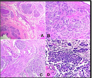
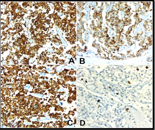
Extrahepatic bile duct Neuroendocrine Tumour (NET) is uncommon, and hence a preoperative diagnosis is rare. The definite diagnosis depends on post-operative histopathology examination and immunohistochemistry studies. Complete excision of the lesion is considered as a curative treatment. We report a case of 39 year old male who presented with obstructive jaundice and imaging suggested Cholangiocarcinoma involving the hepatic ducts. He underwent a right hepatectomy and cholecystectomy with hepatoduodenal lymphadenectomy. The histopathological examination revealed a Neuroendocrine Tumour.
References
- 1. Noronha YS, Raza AS. Well-differentiated neuroendocrine (carcinoid) Tumours of the extra hepatic biliary ducts. Arch Pathol Lab Med 2010; 134 (7): 1075–1079.
- 2. T. Colak, Carcinoid Tumour of the Common Bile Duct: Report of a Case and a Review of the Literature Carcinoid Tumour of the Common Bile Duct: Report of a Case and a Review of, 2006, pp. 1–5, https://doi.org/10.1080/ 00015458.2006.11679850 (February).
- 3. A. Jethava, V. Muralidharan, T. Mesologites, E. Stoica-Mustafa, C.A. Dasanu, H. Medicine, Case report an unusual presentation of a carcinoid tumour of the common bile duct JOP. J. Pancreas (online) 2013 Jan 10, 14 (1) (2013) 85–87.
- 4. K. Hosoda, et al., Neuroendocrine tumour of the common bile duct, Surgery 2016;160(2) (2016) 525-526.
- 5. N. Michalopoulos, et al., Neuroendocrine tumours of extrahepatic biliary tract,Pathol. Oncol. Res. 2014 Oct;20(4):765–775.
- 6. Nagtegaal ID, Odze RD, Klimstra D, Paradis V, Rugge M, Schirmacher P, Washington KM, Carneiro F, Cree IA: WHO Classification of Tumours Editorial Board. The 2019 WHO classification of tumours of the digestive system. Histopathology 2020 Jan;76(2):180-188.
- 7. Taj R, Perez S, Jih LJ, Huynh C, Berumen J, Bouvet M. Primary extrahepatic biliary neuroendocrine tumour: a case report. J Surg Case Reports. 2023 May 27;2023(5):1-3.
- 8. Hubert C, Sempoux C, Berquin A, Deprez F, Jamar F, Giqot JF. Bile duct carcinoid tumours: an uncommondisease but with a good prognosis? Hepato-Gastroenterol. 2005; 52: 1042–7.
- 9. Safwan, M., Vij, M., Govil, S. et al. Well-Differentiated Neuroendocrine Tumour of the Extrahepatic Bile Duct: a Case Report with Review of Literature. J GastrointestCanc 47, 93–99 (2016).
Data Sharing Statement
There are no additional data available. All raw data and code are available upon request.
Funding
This research received no funding.
Author Contributions
All authors contributed significantly to the work and approve its publication.
Ethics Declaration
This article does not involve any human or animal subjects, and therefore does not require ethics approval.
Acknowledgements
We would like to express our gratitude to the patients, their families, and all those who have contributed to this study.
Conflicts of Interest
No conflicts of interest in this work.
About this article

Cite this article
Asha Mary George, Anshika Rai, Neha Sharma. Primary Neuroendocrine Tumour of Extrahepatic Bile Duct: A Rarity. Ind J Pathol: Res Pract. 2023;12(4) 143–146.
Licence:
Attribution-Non-commercial 4.0 International (CC BY-NC 4.0)This license
enables reusers to distribute, remix, adapt, and build upon the material in any
medium or format for noncommercial purposes only, and only so long as
attribution is given to the creator.
| Received | Accepted | Published |
|---|---|---|
| October 22, 2023 | November 25, 2023 | December 30, 2023 |
DOI: https://doi.org/10.21088/ijprp.2278.148X.12423.4
Keywords
Extrahepatic bile duct (EHBD)RHDLHDCHDNETSearch for Similar Articles
Similar Articles
- Clinico-Pathological and Immunohistochemical Study of Primary CNS Tumors in a Te...
- The Role of SGLT2 Inhibitors in Cardiovascular Protection Beyond Diabetes: A Co...
- Epidermal Inclusion Cyst Presenting as 4th Ventricle Space Occupying Lesion: A R...
- Silent Retroperitoneal Seminoma Arising from an Occult Undescended Testis: Diagn...
- Scapular Metastasis of Oral Squamous Cell Carcinoma: A Case Report of an Unusual...
Article Level Metrics
Last UpdatedSunday 20 September 2026, 22:02:22 (IST)
3662
Accesses
13
541
00
Citations
NA
NA
NA
Download citation
Article Keywords
Keyword Highlighting
Highlight selected keywords in the article text.
Timeline
| Received | October 22, 2023 |
| Accepted | November 25, 2023 |
| Published | December 30, 2023 |
licence
Attribution-Non-commercial 4.0 International (CC BY-NC 4.0)
This license
enables reusers to distribute, remix, adapt, and build upon the material in any
medium or format for noncommercial purposes only, and only so long as
attribution is given to the creator.