Full Text (PDF)
Indian Journal of Forensic Medicine and Pathology 13(3):p 419-425, July – September 2020. | DOI: https://doi.org/10.21088/ijfmp.0974.3383.13320.9
Original Article
Pathological Study of Percutaneous Image Guided Biopsy of Vertebral and Paravertebral Lesions; Our Experience
Potti Ramya, Chaitra B1 null, Inuganti Venkata Renuka2 null, Kasula Lakshmi3 null, Vaddatti Tejeswini5 null, Baddula Durgaprasad6 null
Author Information
Licence:
Attribution-Non-commercial 4.0 International (CC BY-NC 4.0)This license enables reusers to distribute, remix, adapt, and build upon the material in any medium or format for noncommercial purposes only, and only so long as attribution is given to the creator.
Indian Journal of Forensic Medicine and Pathology 13(3):p 419-425, July – September 2020. | DOI: https://doi.org/10.21088/ijfmp.0974.3383.13320.9
How Cite This Article:
Chaitra B, Renuka IV, Lakshmi K, et al. Pathological Study of Percutaneous Image Guided Biopsy of Vertebral and Paravertebral Lesions; Our Experience. Indian Journal of Forensic Medicine & Pathology. 2020;13(3):425–431.Timeline
Received : July 02, 2020
Accepted : July 20, 2020
Published : August 30, 2020
Abstract
Background: The presence of a spinal lesion whether symptomatic or not, presents a diagnostic challenge and is always a cause for concern. In Indian population common spinal pathologies include tuberculosis and malignancy. We aim to study the spectrum of vertebral and paravertebral lesions, analyse with regard to age, sex, site, pathological diagnosis and evaluate diagnostic utility of percutaneous image guided biopsy.
Methods: This was an Institutional Ethics Board approved retrospective study conducted in the Department of Pathology on vertebral and paravertebral biopsy specimens received from January 2014 to September 2019.
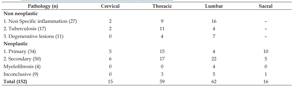
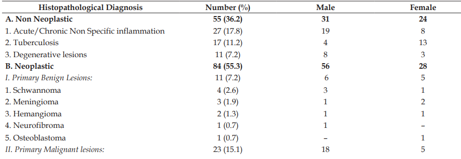
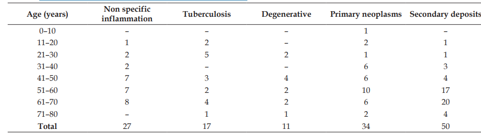
Results: A total of 152 cases of vertebral and paravertebral lesions were reviewed with age range of 2–80 years and majority of 42 cases among 61–70 years age group. Male predominance with male to female ratio of 1.62:1 was noted. Out of 152 cases, a majority of 62 cases were in lumbar region followed by 59 cases in thoracic region. Paravertebral involvement was noted in 24 cases. Among the 152 cases, 55 cases (36.2%) were non-neoplastic lesions, 84 cases (55.3%) were neoplastic. Among neoplastic lesions secondary deposits (60%) were common followed by primary malignancy (27%) and benign tumours (13%). Most common among, metastasis was Adenocarcinoma; primary malignancy was multiple myeloma; and benign tumour was schwannoma. Tuberculosis was seen in 17 cases and majority involving the thoracic segments.
Conclusion: CT guided biopsy is a valuable tool for evaluation of vertebral and paravertebral lesions. Metastatic lesions were common followed by multiple myeloma and schwanoma. Tuberculosis was second common among non neoplastic lesions.
References
- 1. Pant I, Chaturvedi S. Spectrum of histopathology in spinal lesions. Astrocyte. 2016;2(4):187–199.
- 2. Kang M, Gupta S, Khandelwal N, Shankar S, Gulati M, Suri S. CT-Guided Fine-Needle Aspiration Biopsy of Spinal Lesions. Acta Radiologica. 1999;40(5):474–478.
- 3. Aithala JP. Role of Percutaneous Image Guided Biopsy in Spinal Lesions: Adequacy and Correlation with MRI Findings. Journal of Clinical and Diagnostic Research. 2016;10(11):TC11–TC15.
- 4. Gul SB, Polat AV, Bekci T, Selcuk MB. Accuracy of Percutaneous CT-Guided Spine Biopsy and Determinants of Biopsy Success. Journal of the Belgian Society of Radiology. 2016;100(1):58.
- 5. Dikondwar AR, Dani AA. Spinal space occupying lesions - Pathologic spectrum. Journal of Medical Sciences and Health. 2016;2(3):24–29.
- 6. Gadgil NM, Chaudhari CS, Margam SR, et al. A clinicopathological study of lesions of spinal cord and its coverings: A tertiary care hospital experience. Annals of Pathology and Laboratory Medicine. 2016;3(3):148–156.
- 7. Hirano K, Imagama S, Sato K, et al. Primary spinal cord tumors: review of 678 surgically treated patients in Japan. A multicenter study. European Spine Journal. 2012;21(10):2019–2026.
- 8. Arora RK, Kumar R. Spinal tumors: Trends from Northern India. Asian Journal of Neurosurgery. 2015;10(4):291–297.
- 9. Moein P, Behnamfar O, Khalighinejad N, et al. A 12 year epidemiologic study on primary spinal cord tumors in Isfahan, Iran. Journal of Research in Medical Sciences. 2013;18(3):17–21.
- 10. Debnath H, Rahman MZ, Uddin MK, et al. Types and outcomes of spinal cord tumors: A study of 30 cases attending the Department of Neurosurgery in Chittagong Medical College Hospital, Chittagong. Journal of Chittagong Medical College Teachers' Association. 2007;18(1):11–15.
- 11. Feroz I, Makhdoomi RH, Khursheed N, Shaheen F, Shah P. Utility of computed tomography-guided biopsy in evaluation of metastatic spinal lesions. Asian Journal of Neurosurgery. 2018;13(3):577–584.
- 12. Jobanputra GP, Parikh UR, Goswami HM. Histopathological study of spinal tumors. International Journal of Current Research and Review. 2016;8(11):1–8.
- 13. Ciftdemir M, Kaya M, Selcuk E, Yalniz E. Tumors of the spine. World Journal of Orthopedics. 2016;7(2):109–116.
- 14. Schellinger KA, Propp JM, Villano JL, McCarthy BJ. Descriptive epidemiology of primary spinal cord tumors. Journal of Neuro-Oncology. 2008;87(2):173–179.
- 15. Gasbarrini A, Cappuccio M, Mirabile L, et al. Spinal metastases: Treatment evaluation algorithm. European Review for Medical and Pharmacological Sciences. 2004;8(6):265–274.
- 16. Bhat AR, Kirmani AR, Wani MA, Bhat MH. Incidence, histopathology, and surgical outcome of tumors of spinal cord, nerve roots, meninges, and vertebral column - Data based on single institutional (Sher-i-Kashmir Institute of Medical Sciences) experience. Journal of Neurosciences in Rural Practice. 2016;7(3):381–391.
- 17. Jain AK, Singh S, Sinha S, Dhammi IK, Kumar S. Intraspinal tubercular granuloma - an analysis of 17 cases. Indian Journal of Orthopaedics. 2003;37:12–13.
- 18. Govada N, Chowdary KR, Jeshtadi A, Gollapalli S, Dara K. Spinal nerve sheath tumors: Analysis of 20 cases with review of literature. International Journal of Research in Health Sciences. 2014;2(1):140–145.
- 19. Tilva BV, Naik KR, Saroja AO, Ghorpade RS. Spinal intramedullary tuberculoma: A rare cause of paraparesis. Journal of the Scientific Society. 2015;42(2):123–125.
- 20. Koeller KK, Rosenblum RS, Morrison AL. Neoplasms of the Spinal Cord and Filum Terminale: Radiologic-Pathologic Correlation. RadioGraphics. 2000;20(6):1721–1749.
- 21. Celli P, Trillò G, Ferrante L. Spinal extradural schwannoma. Journal of Neurosurgery: Spine. 2005;2(4):447–456.
Data Sharing Statement
There are no additional data available. All raw data and code are available upon request.
Funding
This research received no funding.
Author Contributions
Whether all authors contributed significantly to the work and approve its publication.
Ethics Declaration
This article does not involve any human or animal subjects, and therefore does not require ethics approval.
Acknowledgements
We would like to express our gratitude to the patients, their families, and all those who have contributed to this study.
Conflicts of Interest
The authors report no conflicts of interest in this work.
About this article
Cite this article
Chaitra B, Renuka IV, Lakshmi K, et al. Pathological Study of Percutaneous Image Guided Biopsy of Vertebral and Paravertebral Lesions; Our Experience. Indian Journal of Forensic Medicine & Pathology. 2020;13(3):425–431.
Licence:
Attribution-Non-commercial 4.0 International (CC BY-NC 4.0)This license enables reusers to distribute, remix, adapt, and build upon the material in any medium or format for noncommercial purposes only, and only so long as attribution is given to the creator.
| Received | Accepted | Published |
|---|---|---|
| July 02, 2020 | July 20, 2020 | August 30, 2020 |
DOI: https://doi.org/10.21088/ijfmp.0974.3383.13320.9
Keywords
Image guided biopsyVertebral lesionsSearch for Similar Articles
Similar Articles
- Pesticide Contamination in Indian Agricultural and Residential Areas: A Compara...
- Forensic Age Estimation using CBCT-Derived Mandibular Morphometrics: A Comparat...
- A 2 Years Retrospective Study of the Spectrum of Poisoning in a Tertiary Care C...
- Knowledge and Attitude of MBBS Students Regarding Post Mortem Examination: A Cr...
- Sudden Death in a 7-year-old Child Due to Neoplasm: A Case Report
Article Level Metrics
Last UpdatedWednesday 08 July 2026, 04:31:16 (IST)
7722
Accesses
4
2137
00
Citations
NA
NA
NA
Download citation
Article Keywords
Keyword Highlighting
Highlight selected keywords in the article text.
Timeline
| Received | July 02, 2020 |
| Accepted | July 20, 2020 |
| Published | August 30, 2020 |
licence
Attribution-Non-commercial 4.0 International (CC BY-NC 4.0)
This license enables reusers to distribute, remix, adapt, and build upon the material in any medium or format for noncommercial purposes only, and only so long as attribution is given to the creator.