Full Text (PDF)
Indian Journal of Forensic Medicine and Pathology 13(3):p 411-417, July – September 2020. | DOI: https://doi.org/10.21088/ijfmp.0974.3383.13320.8
Original Article
Histomorphological Analysis of Renal Lesions in Autopsy Specimens
Clement Wilfred D Associate Professor, Department of Pathology, M S Ramaiah Medical College and Hospitals, MSRIT Post, MSR Nagar, Bangalore 560054, India, Likitha SR, Rashmi K null, Usha M null, Viswanath Mysorekar
Author Information
Licence:
Attribution-Non-commercial 4.0 International (CC BY-NC 4.0)This license enables reusers to distribute, remix, adapt, and build upon the material in any medium or format for noncommercial purposes only, and only so long as attribution is given to the creator.
Indian Journal of Forensic Medicine and Pathology 13(3):p 411-417, July – September 2020. | DOI: https://doi.org/10.21088/ijfmp.0974.3383.13320.8
How Cite This Article:
Likitha SR, Wilfred CD, Rashmi K, et al. Histomorphological Analysis of Renal Lesions in Autopsy Specimens. Indian Journal of Forensic Medicine & Pathology. 2020;13(3):411–417.Timeline
Received : July 02, 2020
Accepted : July 02, 2020
Published : August 30, 2020
Abstract
Background: Autopsy is crucial to identify asymptomatic and often undiagnosed lesions. Renal diseases have high morbidity and mortality and the information provided by morphological evaluation of renal autopsy specimens is crucial to prevent renal disease that tends to be asymptomatic. Hence this study was undertaken to analyze the morphological features of kidney lesions in autopsy specimens.
Materials and methods: The study was conducted on renal specimens from routine autopsies, over duration of five years, at a south Indian tertiary health care center. The specimens were evaluated morphologically and histological sections were interpreted and classified into glomerular lesions, nonglomerular lesions and normal findings.
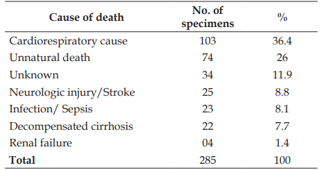
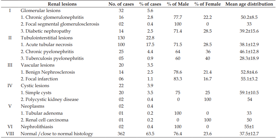
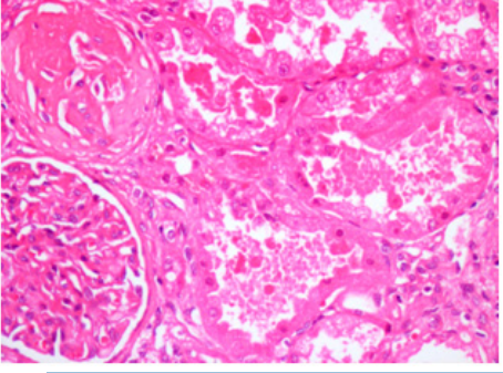
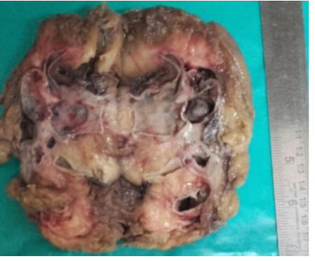
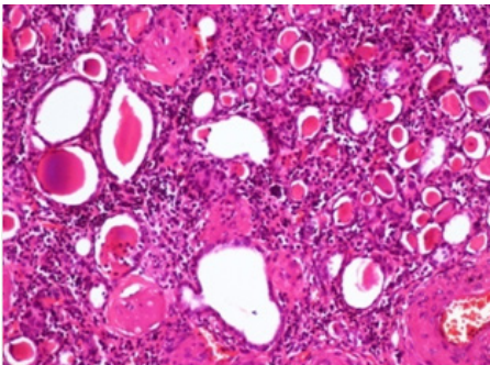
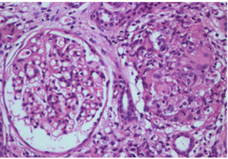
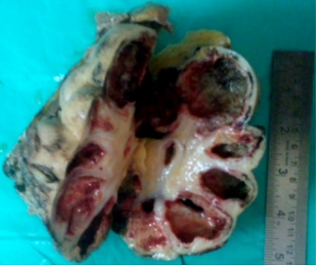
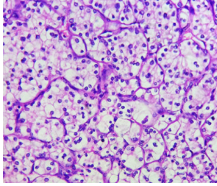
Results: A total of 570 specimens from 285 autopsies, with mean age of 38.8 years and male: female ratio of 3.4:1, were included in the study. The major cause of death was cardiorespiratory failure (36.4%). Normal histology was identified in 63.5% of the kidneys. Tubulointerstitial, glomerular, cystic, vascular and neoplastic lesions were identified in 22.8%, 5.6%, 3.9%, 3.5% and 0.4% of the kidneys, respectively.
Conclusion: The frequency of renal lesions encountered in autopsies was 36.5%. Nonglomerular lesions outnumbered gomerular lesions with male predominance. The commonest tubulointerstitial lesion was acute tubular necrosis. Chronic glomerulonephritis, benign nephrosclerosis and simple renal cysts were the major glomerular, vascular and cystic lesions identified, respectively. Thus, through autopsy, varied preventable renal lesions that tend to be asymptomati
References
- 1. Sandhu VK, Puri A, Singh N. The Histomorphological Spectrum of Renal Lesions in an Autopsy Study. Annals of Pathology and Laboratory Medicine. 2017;4(4):A410–414.
- 2. Patel S, Rajalakshmi BR, Manjunath GV. Histopathologic Findings in Autopsies with Emphasis on Interesting and Incidental Findings - A Pathologist’s Perspective. Journal of Clinical and Diagnostic Research. 2016;10(11):EC08–EC12.
- 3. Chethan K, Shashikala P, Deepti P, Kavita GU. Histomorphological Spectrum of Kidney Lesions in Nephrectomies and Autopsies. International Journal of Scientific Research. 2016;5(6):366–367.
- 4. Henriksen KJ. Assessment of kidneys in adult autopsies. Diagnostic Histopathology. 2017;23(3):117–125.
- 5. Schiffrin EL, Lipman ML, Mann JF. Chronic kidney disease: effects on the cardiovascular system. Circulation. 2007;116(1):85–97.
- 6. Levey AS, Atkins R, Coresh J, Cohen EP, Collins AJ, Eckardt KU, et al. Chronic kidney disease as a global public health problem: approaches and initiatives - a position statement from Kidney Disease Improving Global Outcomes. Kidney International. 2007;72(2):247–259.
- 7. Perrone ME, Chang A, Henriksen KJ. Medical renal diseases are frequent but often unrecognized in adult autopsies. Modern Pathology. 2018;31:365–373.
- 8. Yadav SNS, Bhattacharya AB. Histomorphological Study of Kidney Lesions in Autopsy – An Original Eight Year Study. IOSR Journal of Dental and Medical Sciences. 2019;18(3):50–54.
- 9. Pandian JR, Laishram RS, Kumar LD, Phuritsabam P, Debnath K. Autopsy review of sudden deaths in a tertiary hospital of northeastern India. Journal of Medical Society. 2014;28(3):145–148.
- 10. Kaur A, Bodal VK, Garg P, Aggarwal A. Histopathological Spectrum of Kidney lesions in autopsy - A study of 100 cases. Journal of Medical Science and Clinical Research (JMSCR). 2018;6(2):962–966.
- 11. Usta U, Tastekin E, Isler E, Kutlu AK, Puyan FO. Histopathological and immune alterations in autopsied kidneys. Saudi Medical Journal. 2014;35(11):1331–1338.
- 12. Hailemariam S, Walder M, Burger HR, Cathomas G, Mihatsch M, Binswanger U, et al. Renal pathology and premortem clinical presentation of Caucasian patients with AIDS: An autopsy study from the era prior to antiretroviral therapy. Swiss Medical Weekly. 2001;131:412–417.
- 13. Kocovski L, Duflou J. Can renal acute tubular necrosis be differentiated from autolysis at autopsy? Journal of Forensic Sciences. 2009;54(2):439–442.
- 14. Eastwood JB, Corbishley CM, Grange JM. Tuberculosis and the kidney. Journal of the American Society of Nephrology. 2001;12(6):1307–1314.
- 15. Alpers CE, Chang A. The Kidney. In: Kumar V, Abbas AK, Aster JC, editors. Robbins & Cotran Pathologic Basis of Disease. 8th ed. Philadelphia: Elsevier/Saunders; 2015. p. 945–950.
Data Sharing Statement
There are no additional data available. All raw data and code are available upon request.
Funding
This research received no funding.
Author Contributions
Whether all authors contributed significantly to the work and approve its publication.
Ethics Declaration
This article does not involve any human or animal subjects, and therefore does not require ethics approval.
Acknowledgements
We would like to express our gratitude to the patients, their families, and all those who have contributed to this study.
Conflicts of Interest
The authors report no conflicts of interest in this work.
About this article
Cite this article
Likitha SR, Wilfred CD, Rashmi K, et al. Histomorphological Analysis of Renal Lesions in Autopsy Specimens. Indian Journal of Forensic Medicine & Pathology. 2020;13(3):411–417.
Licence:
Attribution-Non-commercial 4.0 International (CC BY-NC 4.0)This license enables reusers to distribute, remix, adapt, and build upon the material in any medium or format for noncommercial purposes only, and only so long as attribution is given to the creator.
| Received | Accepted | Published |
|---|---|---|
| July 02, 2020 | July 02, 2020 | August 30, 2020 |
DOI: https://doi.org/10.21088/ijfmp.0974.3383.13320.8
Keywords
AutopsyGlomerular lesionsNonglomerular lesionsRenal lesionsRenal specimensSearch for Similar Articles
Similar Articles
- Pesticide Contamination in Indian Agricultural and Residential Areas: A Compara...
- Forensic Age Estimation using CBCT-Derived Mandibular Morphometrics: A Comparat...
- A 2 Years Retrospective Study of the Spectrum of Poisoning in a Tertiary Care C...
- Knowledge and Attitude of MBBS Students Regarding Post Mortem Examination: A Cr...
- Sudden Death in a 7-year-old Child Due to Neoplasm: A Case Report
Article Level Metrics
Last UpdatedWednesday 08 July 2026, 04:27:49 (IST)
7722
Accesses
3
2137
00
Citations
NA
NA
NA
Download citation
Article Keywords
Keyword Highlighting
Highlight selected keywords in the article text.
Timeline
| Received | July 02, 2020 |
| Accepted | July 02, 2020 |
| Published | August 30, 2020 |
licence
Attribution-Non-commercial 4.0 International (CC BY-NC 4.0)
This license enables reusers to distribute, remix, adapt, and build upon the material in any medium or format for noncommercial purposes only, and only so long as attribution is given to the creator.