Full Text (PDF)
Indian Journal of Pathology: Research and Practice 13(4):p 111-115, Oct. Dec. 2024. | DOI: https://doi.org/10.21088/ijprp.2278.148X.13324.4
Case Report
Nevus Lipomatosus Cutaneous Superficialis: A Rare Occurrence
Jyoti Verma, Jyotsna N. Bharti, Rajesh K. Jarang, Rajashekar Mohan
Author Information
Licence:
Attribution-Non-commercial 4.0 International (CC BY-NC 4.0)This license enables reusers to distribute, remix, adapt, and build upon the material in any medium or format for noncommercial purposes only, and only so long as attribution is given to the creator
Indian Journal of Pathology: Research and Practice 13(4):p 111-115, Oct. Dec. 2024. | DOI: https://doi.org/10.21088/ijprp.2278.148X.13324.4
How Cite This Article:
Jyoti Verma, Jyotsna N. Bharti, Rajesh K. Jarang et al., Nevus Lipomatosus Cutaneous Superficialis: A Rare Occurrence. Ind Jr of Path: Res and Practice 2024;13(3)111-115.Timeline
Received : January 14, 2024
Accepted : February 10, 2024
Published : December 30, 2024
Abstract
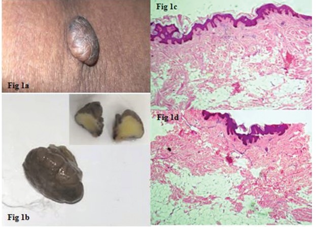
Nevus lipomatosus cutaneous superficialis (NLCS) is a rare cutaneous hamartoma characterized by the ectopic presence of mature adipocytes in papillary and reticular dermis without connection to the subcutis. It usually has a predilection for buttocks, upper thigh and back. It presents itself as skin to yellow colored discrete or confluent papulo-nodular soft, fleshy sessile mass and has two clinical forms described in the literature, classical and solitary types. The classical form is usually composed of multiple and grouped skin-colored, pedunculated nodules. A second and more rare form is characterized by a solitary dome-shaped sessile papule or nodule. Microscopically it presents with the fat deposition in the dermis and is considered to be the consequence of the degenerative changes in the connective tissues. Its pathogenesis is unknown; however, several theories have been proposed. Some attribute it to the adipocyte metaplasia during the degenerative course and some to the development displacement of adipose tissue while others, as a consequence of maturation of adipocytes from mononuclear cells differentiation into lipoblasts in the perivascular zone. Histopathology shows clusters of ectopic presence of adipose fat among the desiccating collagen fibres in the dermis without any connection with the subcutaneous fat. Herein, we present a case of solitary NLCS over left flank associated with left upper thigh acrochordon in a 30 years female.
References
- 1. Jain A, Sharma A, Sharda R, Aggarwal C. Nevus lipomatosus cutaneous superficialis: A rare hamartoma. Indian J Surg Oncol. 2020; 11:147-9.
- 2. Hoffman E, Zurhelle E. Ubre einen naevus lipomatosus cutaneous superficialis der linken glutaalgegend. Arch Dermatol. 1921; 150:327-33.
- 3. Alotaibi H, Alsaif F, Alali A et al.,Nevus Lipomatosis Cutaneous Superficialis: A Single-Center Case Series of 5 Patients. Case Rep Dermatol 2018; 10:138–144.
- 4. Dhamija A, Mehedra A, D’Souza P, Meena RS. Nevus lipomatosus cutaneous superficialis: An unusual presentation. Indian Dermatol Online J. 2012; 3:196-98
- 5. Ragsdale BD. Tumors with fatty, muscular, osseous, and/or cartilaginous differentiation. In: Elder DE, Elenitsas R, Jhonson BL, Murphy GF, Xu X, editors. Lever’s histopathology of the skin. 10th ed. Wolters Kluwer-Lippincott Williams & Wilkins; 2009.
- 6. Goucha S, Khaled A, Zéglaoui F, Rammeh S, Zermani R, Fazaa B. Nevus lipomatosus cutaneous superficialis: report of eight cases. Dermatol Ther (Heidelb). 2011 Dec; 1(2):25–30.
- 7. Triki S, Mekni A, Haouet S, Mokni M, Kchir N, Ben Osman Dhahri A et al. [Nevus lipomatosus cutaneous superficialis: a clinico-pathological study of 13 cases]. Tunis Med. 2006 Dec; 84(12):800–2. French.
- 8. Meenu Pujani, Monisha Choudhury, Taru Garg et al., Nevus lipomatosus superficialis: A rare cutaneous hamartoma. Indian Dermatol Online J. 2014 Jan-Mar; 5(1): 109–110.
- 9. Leung AK, Barankin B. Nevus lipomatosus superficialis on the left proximal arm. Case Dermatol Med. 2017; 10:1155.
- 10. Bhushan P, Thatte SS, Sing A. Nevus lipomatosus cutaneous superficialis: A report of two cases. Indian J Dermatol. 2016; 61:123.
- 11. Bancalari E, Martinez-Sanchez D, Tardio JC. Nevus lipomatosus superficialis with a folliculosebaceous component: report of 2 cases. Patholog Res Int. 2011 Apr; 2011:105973.
- 12. Lane JE, Clark E, Marzec T. Nevus lipomatosus cutaneus superficialis. Pediatr Dermatol. 2003 JulAug; 20(4):313–4.
- 13. Sardana K, Bansal S, Garg VK, Khurana N. Treatment of nevus lipomatosus cutaneous superficialis with CO2 laser. J Cosmet Dermat. 2017; 16:333-5.
Data Sharing Statement
There are no additional data available. All raw data and code are available upon request.
Funding
This research received no funding.
Author Contributions
All authors contributed significantly to the work and approve its publication.
Ethics Declaration
This article does not involve any human or animal subjects, and therefore does not require ethics approval.
Acknowledgements
We would like to express our gratitude to the patients, their families, and all those who have contributed to this study.
Conflicts of Interest
No conflicts of interest in this work.
About this article

Cite this article
Jyoti Verma, Jyotsna N. Bharti, Rajesh K. Jarang et al., Nevus Lipomatosus Cutaneous Superficialis: A Rare Occurrence. Ind Jr of Path: Res and Practice 2024;13(3)111-115.
Licence:
Attribution-Non-commercial 4.0 International (CC BY-NC 4.0)This license enables reusers to distribute, remix, adapt, and build upon the material in any medium or format for noncommercial purposes only, and only so long as attribution is given to the creator
| Received | Accepted | Published |
|---|---|---|
| January 14, 2024 | February 10, 2024 | December 30, 2024 |
DOI: https://doi.org/10.21088/ijprp.2278.148X.13324.4
Keywords
Nevus lipomatosus cutaneous superficialisHamartomaEctopicAdipocytesSearch for Similar Articles
Similar Articles
- Clinico-Pathological and Immunohistochemical Study of Primary CNS Tumors in a Te...
- The Role of SGLT2 Inhibitors in Cardiovascular Protection Beyond Diabetes: A Co...
- Epidermal Inclusion Cyst Presenting as 4th Ventricle Space Occupying Lesion: A R...
- Silent Retroperitoneal Seminoma Arising from an Occult Undescended Testis: Diagn...
- Scapular Metastasis of Oral Squamous Cell Carcinoma: A Case Report of an Unusual...
Article Level Metrics
Last UpdatedTuesday 22 September 2026, 02:39:45 (IST)
3670
Accesses
10
541
00
Citations
NA
NA
NA
Download citation
Article Keywords
Keyword Highlighting
Highlight selected keywords in the article text.
Timeline
| Received | January 14, 2024 |
| Accepted | February 10, 2024 |
| Published | December 30, 2024 |
licence
Attribution-Non-commercial 4.0 International (CC BY-NC 4.0)
This license enables reusers to distribute, remix, adapt, and build upon the material in any medium or format for noncommercial purposes only, and only so long as attribution is given to the creator