Full Text (PDF)
Indian Journal of Pathology: Research and Practice 12(1):p 29-33, January-March 2023. | DOI: 10.21088/ijprp.2278.148X.12123.4
Case Report
Invasive Mucormycosis in an Adolescent Female with Type-I Diabetes Mellitus
Sunil V Jagtap, Shubham S Jagtap, Devika Suresh Borade, Gauri Avinash Patil, Shuchita Gaur
Author Information
Licence:
Attribution-Non-commercial 4.0 International (CC BY-NC 4.0)This license enables reusers to distribute, remix, adapt, and build upon the material in any medium or format for noncommercial purposes only, and only so long as attribution is given to the creator.
Indian Journal of Pathology: Research and Practice 12(1):p 29-33, January-March 2023. | DOI: 10.21088/ijprp.2278.148X.12123.4
How Cite This Article:
Jagtap SV, Jagtap SS, Borade DS, et al. Invasive mucormycosis in an adolescent female with type-I diabetes mellitus. Indian J Pathol Res Pract. 2023;12(1):29–33.Timeline
Received : November 23, 2022
Accepted : December 28, 2022
Published : January 30, 2023
Abstract
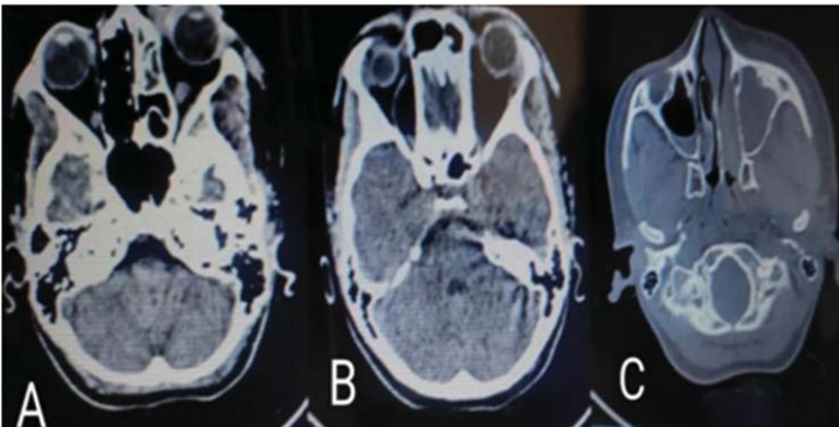
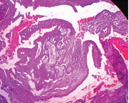
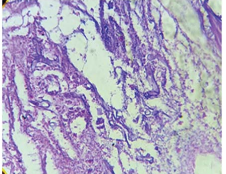
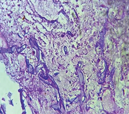
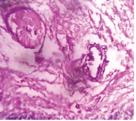
Mucormycosis is aggressive and potentially fatal in patients, particularly among diabetic patients suffering from ketoacidosis. A 16-year female patient came to the medical department with a complaint of fever for 15 days, cough and breathlessness, chest pain, and palpitation for 10 days also had left orbital and facial swelling for 3 days. The patient is a known case of type 2 diabetes mellitus. On CT Scan PNS (Plain) showed a soft tissue thickening causing complete opacification of the left maxillary sinus extending to the left frontal, left ethmoid, and left he misphenoid sinuses. The treatment of debridement of necrotic tissue from the left maxillary sinus was done. On histopathology diagnosed as invasive mucormycosis. We are presenting this case of mucormycosis in diabetic patients suffering from ketoacidosis for its clinical, radiological, and histopathological findings and management of the disease. Early diagnosis and prompt treatment significantly improve survival and decreases morbidity.
References
- 1. Auluck A. Maxillary necrosis by mucormycosis. A case report and literature review. Med Oral Patol Oral Cir Bucal. 2007;12(5):E360–4.
- 2. Roden MM, Zaoutis TE, Buchanan WL, Knudsen TA, Sarkisova TA, Schaufele RL, et al. Epidemiology and outcome of zygomycosis: a review of 929 reported cases. Clin Infect Dis. 2005;41(5):634–53.
- 3. Petrikkos G, Skiada A, Lortholary O, Roilides E, Walsh TJ, Kontoyiannis DP. Epidemiology and clinical manifestations of mucormycosis. Clin Infect Dis. 2012;54(Suppl 1):S23–34.
- 4. Sunil VJ, Swati SJ, Atul H, Snigdha AV, Divya B, Shubhang M. COVID-19 associated mucormycosis (CAM): a clinico-histopathological study at a tertiary care centre. Indian J Pathol Res Pract. 2022;11(1):19–26.
- 5. Jeong W, Keighley C, Wolfe R, Lee WL, Slavin MA, Kong DCM, et al. The epidemiology and clinical manifestations of mucormycosis: a systematic review and meta-analysis of case reports. Clin Microbiol Infect. 2019;25:26–34.
- 6. Jagtap SV, Jagtap SS, Nagar V, Varshney K. Invasive mucormycosis in post COVID-19 infection: case report with review. IP Arch Cytol Histopathol Res. 2021;6(2):135–9.
- 7. Karadeniz Uğurlu Ş, Selim S, Kopar A, Songu M. Rhino-orbital mucormycosis: clinical findings and treatment outcomes of four cases. Turk J Ophthalmol. 2015;45(4):169–74.
- 8. Bavikar P, Mehta V. Rhino-orbital-cerebral mucormycosis: a fatal complication of uncontrolled diabetes mellitus. Cureus. 2017;9(11):e1841.
- 9. Jagtap SV, Hulwan A, Vartak S, et al. Co-infection of mucormycosis and actinomycosis in COVID-19 infection. Int J Health Sci Res. 2021;11(8):127–30.
- 10. Galletti B, Gazia F, Galletti C, Perani F, Ciodaro F, Freni F, et al. Rhinocerebral mucormycosis with dissemination to pontine area in a diabetic patient: treatment and management. Clin Case Rep. 2019;7(7):1382–7.
- 11. Bhansali A, Bhadada S, Sharma A, et al. Presentation and outcome of rhino-orbital-cerebral mucormycosis in patients with diabetes. Postgrad Med J. 2004;80:670–4.
Data Sharing Statement
There are no additional data available. All raw data and code are available upon request.
Funding
This research received no funding.
Author Contributions
Whether all authors contributed significantly to the work and approve its publication.
Ethics Declaration
This article does not involve any human or animal subjects, and therefore does not require ethics approval.
Acknowledgements
We would like to express our gratitude to the patients, their families, and all those who have contributed to this study.
Conflicts of Interest
The authors report no conflicts of interest in this work.
About this article

Cite this article
Jagtap SV, Jagtap SS, Borade DS, et al. Invasive mucormycosis in an adolescent female with type-I diabetes mellitus. Indian J Pathol Res Pract. 2023;12(1):29–33.
Licence:
Attribution-Non-commercial 4.0 International (CC BY-NC 4.0)This license enables reusers to distribute, remix, adapt, and build upon the material in any medium or format for noncommercial purposes only, and only so long as attribution is given to the creator.
| Received | Accepted | Published |
|---|---|---|
| November 23, 2022 | December 28, 2022 | January 30, 2023 |
DOI: 10.21088/ijprp.2278.148X.12123.4
Keywords
Rhinoorbital Infections.MucormycosisDiabetic KetoacidosisOpportunistic InfectionSearch for Similar Articles
Similar Articles
- Recurrent and Subacute Intestinal Obstruction and Ileocecal Stricture due to End...
- Colarado Microdissection Needle: A Review
- Role of Immunohistochemical Typing in Pancreaticobiliary and Intestinal Type Dif...
- Correlation Study of Fine Needle Aspiration Cytology of Thyroid Lesions With Thy...
- Evaluation of Thrombocytopenia with Special References to Platelet Indices in Fi...
Article Level Metrics
Last UpdatedThursday 09 July 2026, 01:35:06 (IST)
1738
Accesses
11
315
00
Citations
NA
NA
NA
Download citation
Article Keywords
Keyword Highlighting
Highlight selected keywords in the article text.
Timeline
| Received | November 23, 2022 |
| Accepted | December 28, 2022 |
| Published | January 30, 2023 |
licence
Attribution-Non-commercial 4.0 International (CC BY-NC 4.0)
This license enables reusers to distribute, remix, adapt, and build upon the material in any medium or format for noncommercial purposes only, and only so long as attribution is given to the creator.