Full Text (PDF)
Indian Journal of Forensic Medicine and Pathology 13(1):p 19-31, January-March 2020. | DOI: https://doi.org/10.21088/ijfmp.0974.3383.13120.3
Original Article
Histopathological Spectrum of Ovarian Tumors: A 3-Year Retrospective Study in a Tertiary Care Centre in Southern India
Swati Sharma, Anugnya P Ranjoalkar, Manna Valiathan, Kanthilatha Pai, Muralidhar Pai
Author Information
Licence:
Attribution-Non-commercial 4.0 International (CC BY-NC 4.0)This license enables reusers to distribute, remix, adapt, and build upon the material in any medium or format for noncommercial purposes only, and only so long as attribution is given to the creator
Indian Journal of Forensic Medicine and Pathology 13(1):p 19-31, January-March 2020. | DOI: https://doi.org/10.21088/ijfmp.0974.3383.13120.3
How Cite This Article:
Anugnya P Ranjoalkar, Swati Sharma, Manna Valiathan et al. Histopathological Spectrum of Ovarian Tumors: A 3-YearRetrospective Study in a Tertiary Care Centre in Southern India. Indian J. Forensic Med Pathol. 2020;13(1):19-31.Timeline
Received : January 02, 2020
Accepted : February 02, 2020
Published : March 30, 2020
Abstract
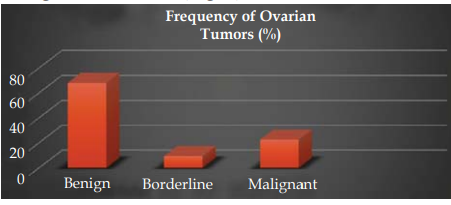
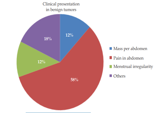
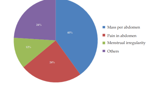
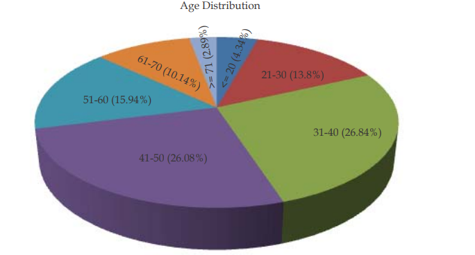
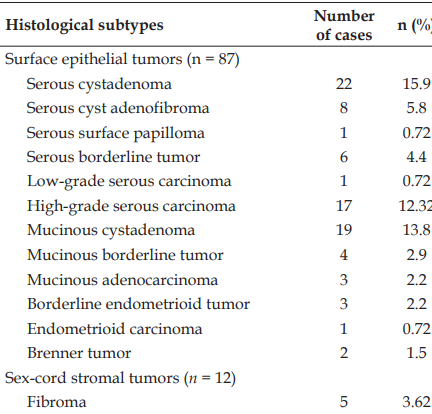
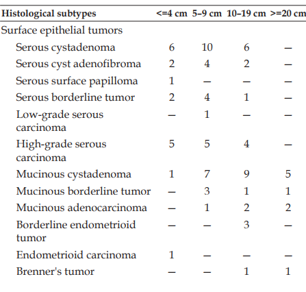
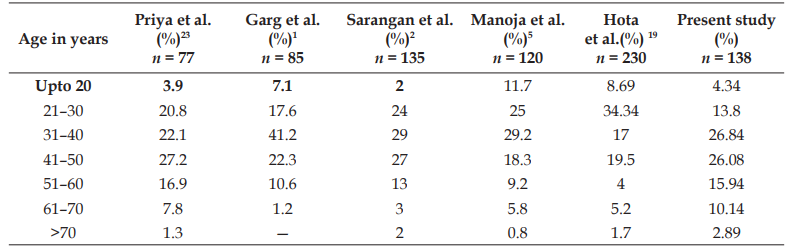
Introduction: Ovarian tumors are a group of diverse neoplasms with a varied clinical, morphological and histological feature. The varied anatomy, histogenesis and its peculiar physiology including the cyclical changes from puberty to menopause give rise to number of cell types, each of which may give rise to tumors. Materials and methods: A 3-year retrospective study of histologically proven ovarian neoplasms where the tumors were classified according to World Health Organization (WHO) 2014 classification and their clinical and histopathologic parameters were analyzed. Results: Of all 138 ovarian tumors studied, 94 (68.12%) were benign, 13 (9.42%) borderline and 31 (22.5%) were malignant in nature. Benign tumors chiefly presented with abdominal pain with median age of 39. Mature cystic teratoma was found to be the most common benign tumor. Borderline tumors presented at a median age of 37. Borderline serous and mucinous tumors (30.76%) were the most common borderline tumors. Malignant tumors presented frequently with abdominal mass and at median age of 48. According to WHO classification of tumors based on cell of origin, surface epithelial tumor were the most common ovarian neoplasms, accounting for 63.04% cases, followed by germ cell tumor (24%) and sex-cord stromal tumors (8.7%). Conclusion: Surface epithelial tumors were the most common histopathological subtype of ovarian tumors. Benign and borderline tumors were predominantly found in reproductive age, whereas malignant tumors were seen in perimenopausal and postmenopausal women. Since the prognosis, therapeutic strategies including multidisciplinary approach depend primarily on the histopathologic diagnosis, an accurate pathological evaluation and classification is of prime importance. The multidisciplinary approach employed has its own medico legal implications.
Keywords: Neoplasm; Epithelial Cancer; Ovarian
References
- 1. Garg N, Anand AS, Annigeri C. Study of histomorphological spectrum of ovarian tumors. Int J Med Health Res. 2017;3(10):12–20.
- 2. Sarangan A, Kilpauk G, Andal N. Clinicopathological and histological features of ovarian tumor: a study. IOSR J Dent Med Sci. 2017;16(9):56–60.
- 3. Parmar P, Sehgal S, Mathur K, Yadav A. Histopathological study of ovarian tumors in tertiary care center. Int J Med Res Prof. 2017;3(2):96–8.
- 4. Pradhan A, Upreti D. Histopathological patterns of ovarian tumors at BPKIHS. Health Renaissance. 2012;10(2):87–97.
- 5. Manoja V, Pramood M, Jyothi V, et al. Clinicopathological study of ovarian tumors: a 2-year study. Int J Sci Study. 2017;5(3):300–5.
- 6. Pachori G, Meena U, Sunaria R, et al. Histopathological study of ovarian tumors in Ajmer region. Int J Med Sci Public Health. 2016;5(7):1400.
- 7. Farooq F, Noman D, Humayun N, et al. Demographic differentials and histopathological patterns of ovarian masses. Biomedica. 2015;31(2):118–23.
- 8. Parvatala A, Rajendra Prasad J, Rao NB, et al. Study of neoplastic lesions of the ovary. IOSR J Dent Med Sci. 2015;14:92–6.
- 9. Bhagyalakshmi A, Sreelekha A, Sridevi S, et al. Prospective study of histopathological patterns of ovarian tumors in a tertiary care centre. Int J Res Med Sci. 2014;2(2):448.
- 10. Danish F, Khanzada MS, Mirza T, et al. Histomorphological spectrum of ovarian tumors with immunohistochemical analysis of poorly or undifferentiated malignancies. Gomal J Med Sci. 2012;10(2):209–15.
- 11. Gangaraju S, Sarella LK, Gurugubelli S, et al. Scenario of ovarian mass lesions at a teaching hospital in Andhra Pradesh, India. Int J Reprod Contracept Obstet Gynecol. 2015;4(4):982–9.
- 12. Jindal U. Pattern of ovarian neoplasm in rural population: a five-year study from tertiary care hospital. J Evol Med Dent Sci. 2014;3(8):24.
- 13. Vargas AN. Natural history of ovarian cancer. Ecancermedicalscience. 2014;8:465.
- 14. Travis WD, Brambilla E, Muller-Hermelink HK, et al. World Health Organization classification of tumours. 4th ed. Lyon: IARC Press; 2008. p. 8–83.
- 15. Okugawa K, Hirakawa T, Fukushima K, et al. Relationship between age, histological type, and size of ovarian tumors. Int J Gynaecol Obstet. 2001;74(1):45–50.
- 16. Rajagopal L, Ravikumar U. Diagnostic utility of clinicopathological correlation in ovarian tumors: an analysis of 200 cases. Int J Pharm Bio Sci. 2015;6(1):B1054–73.
- 17. Patel AS, Patel JM, Shah KJ. Ovarian tumors - incidence and histopathological spectrum in tertiary care center, Valsad. IAIM. 2018;5(2):84–93.
- 18. Patil RK, Bhandari BJ, Kittur SK, et al. Histomorphological study of ovarian tumors at a tertiary care centre. Ann Pathol Lab Med. 2017;4(6):0–7.
- 19. Hota R, Panda KM, Bhuyan T. Clinical and histopathological correlation of ovarian tumor. IOSR J Dent Med Sci. 2018;17(7):66–71.
- 20. Shen F, Chen S, Gao Y, et al. The prevalence of malignant and borderline ovarian cancer in pre- and post-menopausal Chinese women. Oncotarget. 2017;8(46):80589–94.
- 21. Hawaldar R, Sodani S, Patidar E. Histopathological spectrum of ovarian tumors: a two year retrospective study. Indian J Pathol Oncol. 2017;4(3):450–3.
- 22. Agrawal P, Kulkarni DG, Chakrabarti PR, et al. Clinicopathological spectrum of ovarian tumors: a 5–year experience in a tertiary health care center. J Basic Clin Reprod Sci. 2015;4(2):90–6.
- 23. Priya RP, Sundari KPM, Rani RA. Overview of ovarian masses. Int J Reprod Contracept Obstet Gynecol. 2016;5(11):3770–2.
- 24. Malli M, Vyas B, Gupta S, et al. A histological study of ovarian tumors in different age groups. Int J Med Sci Public Health. 2014;3(3):338.
- 25. Singh S, Saxena V, Lata S, et al. Histopathological evaluation of ovarian tumors. Chemother Res Pract. 2016;2015:435–9.
- 26. Bindal J, Bankey S. Prevalence of ovarian tumors among ovarian mass lesions in Gajra Raja Medical College, Gwalior, India. Int J Reprod Contracept Obstet Gynecol. 2017;6(9):3907–10.
- 27. Jha R, Karki S. Histological pattern of ovarian tumors and their age distribution. Nepal Med Coll J. 2008;10(2):81–5.
- 28. Baru L, Patnaik R, Singh KB. Clinico-pathological study of ovarian tumors. Int J Reprod Contracept Obstet Gynecol. 2017;6(8):3438–44.
- 29. Rajavigneshwari N, Kotasthane DS, Koteeswaran G. Clinicopathological spectrum of ovarian tumors in a tertiary care hospital. J Evol Med Dent Sci. 2017;6(36):2948–52.
- 30. Jain G, Mankar D. Histopathological profile of ovarian tumors: a twelve-year institutional experience. Muller J Med Sci Res. 2015;6(2):107.
- 31. Phukan A, Borgogoi M, Ghosh S. Histopathological spectrum of ovarian tumors: an institutional perspective. Int J Res Med Sci. 2018;6(8):2639–43.
- 32. Atanda TA, Mohammed AZ, Zakari MS. A seven-year histopathological review of malignant ovarian tumors seen in Kano. Trop J Obstet Gynaecol. 2009;26(2).
- 33. Ahmed M, Afroze N, Sabiha M. Morphological pattern of ovarian tumor: experience in a tertiary level hospital. J Bangladesh Coll Physicians Surg. 2018;36(1):5–10.
- 34. Pilli GS, Suneeta KP, Dhanded AV, et al. Ovarian tumors: a study of 282 cases. J Indian Med Assoc. 2002;100:420, 432–4, 447.
- 35. Sheikh S, Bashir H, Farooq S, et al. Histopathological spectrum of ovarian tumors from a referral hospital in Kashmir valley, Jammu and Kashmir, India. Int J Res Med Sci. 2017;5(5):2110–4.
- 36. Mohapatro M, Dash D, Rao ES. A study on clinicopathological spectrum of ovarian tumors in a tertiary care centre. J Evid Based Med Healthc. 2017;4(37):2223–30.
- 37. Akakpo PK, Derkyi-Kwarteng L, Gyasi RK, et al. A pathological and clinical study of 706 primary tumors of the ovary in the largest tertiary hospital in Ghana. J Bangladesh Coll Physicians Surg. 2017;17(1):1–6.
- 38. Clinical Oncological Society of Australia, Cancer Council Australia, National Cancer Control Initiative. Optimising cancer care in Australia [Internet]. Melbourne: NCCI;
- 39. Victorian Government Department of Human Services. Ministerial taskforce for cancer: achievements 2003–2007 [Internet]. Melbourne: DHS;
- 40. Ruhstaller T, Roe H, Thurlimann B, et al. The multidisciplinary meeting: an indispensible aid to communication between different specialities. Eur J Cancer. 2006;42(15):2459–62.
Data Sharing Statement
There are no additional data available. All raw data and code are available upon request.
Funding
This research received no funding.
Author Contributions
Whether all authors contributed significantly to the work and approve its publication.
Ethics Declaration
This article does not involve any human or animal subjects, and therefore does not require ethics approval.
Acknowledgements
We would like to express our gratitude to the patients, their families, and all those who have contributed to this study.
Conflicts of Interest
The authors report no conflicts of interest in this work.
About this article
Cite this article
Anugnya P Ranjoalkar, Swati Sharma, Manna Valiathan et al. Histopathological Spectrum of Ovarian Tumors: A 3-YearRetrospective Study in a Tertiary Care Centre in Southern India. Indian J. Forensic Med Pathol. 2020;13(1):19-31.
Licence:
Attribution-Non-commercial 4.0 International (CC BY-NC 4.0)This license enables reusers to distribute, remix, adapt, and build upon the material in any medium or format for noncommercial purposes only, and only so long as attribution is given to the creator
| Received | Accepted | Published |
|---|---|---|
| January 02, 2020 | February 02, 2020 | March 30, 2020 |
DOI: https://doi.org/10.21088/ijfmp.0974.3383.13120.3
Keywords
NeoplasmEpithelial CancerOvarianSearch for Similar Articles
Similar Articles
- Analysis of Death Due to Pulmonary Embolism, a Case Series
- Suspected Adverse Event Following Immunization with Multisystem Inflammatory Res...
- Drone: A Smart Intelligent Framework Aiding Forensic Investigations
- Assessment of Knowledge on Sexual Assault Forensic Examination among Nurses in I...
- An Analysis of the Medico-Legal Aspects and Trends in Sexual Offense Cases at a...
Article Level Metrics
Last UpdatedThursday 09 July 2026, 08:58:07 (IST)
7829
Accesses
5
2153
00
Citations
NA
NA
NA
Download citation
Article Keywords
Keyword Highlighting
Highlight selected keywords in the article text.
Timeline
| Received | January 02, 2020 |
| Accepted | February 02, 2020 |
| Published | March 30, 2020 |
licence
Attribution-Non-commercial 4.0 International (CC BY-NC 4.0)
This license enables reusers to distribute, remix, adapt, and build upon the material in any medium or format for noncommercial purposes only, and only so long as attribution is given to the creator