Full Text (PDF)
Indian Journal of Forensic Medicine and Pathology 16(2):p 109-114, April–June 2023. | DOI: https://doi.org/10.21088/ijfmp.0974.3383.16223.3
Original Article
Histopathological Features of Skin changes caused by Electrocution: An Autopsy study
Vinodini S, Divya Lakshmi PK, Saranya S
Author Information
Licence:
Attribution-Non-commercial 4.0 International (CC BY-NC 4.0)This license enables reusers to distribute, remix, adapt, and build upon the material in any medium or format for noncommercial purposes only, and only so long as attribution is given to the creator
Indian Journal of Forensic Medicine and Pathology 16(2):p 109-114, April–June 2023. | DOI: https://doi.org/10.21088/ijfmp.0974.3383.16223.3
How Cite This Article:
Divya Lakshmi PK, Vinodini S, Saranya S. Histopathological features of skin changes caused by electrocution: an autopsy study. Indian J Forensic Med Pathol. 2023;16(2):109-114.Timeline
Received : January 25, 2023
Accepted : April 01, 2023
Published : June 30, 2023
Abstract
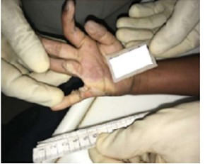
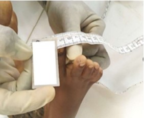
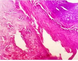
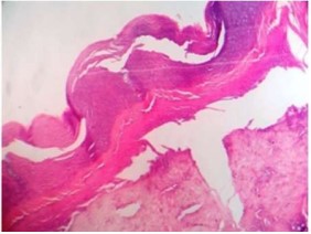
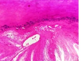
Dependency to electrical equipments and improper safety measures has led to a signifi cant increase in deaths due to electrocution. Confi rming the cause of death in electrocution is one of the biggest challenges. Skin is the most frequently involved tissue in electrocution and histopathology of skin from electric marks aids in proper diagnosis. To study the histomorphological features of epidermis and dermis in skin tissue of entry and exit wounds in electrocution deaths. This is an observational study done over a period of one year. Skin biopsy from medico legal autopsy of non-lightening electrocution deaths obtained from entry or exit wound site were included for the study. Gross and histopathological details were studied and data analyzed. Signifi cant gross features were surface discoloration of skin 16 (61%) and ulceration 05 (20%). Microscopic features were coagulative necrosis 21 (80.7%), dermo-epidermal separation 20 (80.7%), focal ulceration of epithelium 05 (19%), clefting of epidermis (60%), streaming of nuclei (92.3%), elongation of nuclei (92.3%) and microblisters 02 (15.3%). One of the important causes of negative autopsy is electrocution deaths. In such cases dermatopathology is a diagnostic tool coupled with circumstantial evidence. Histological hallmark of electrocution skin injury is epidermal nuclear elongation.
References
- 1. Giri S, Waghmode A, Tumram NK. Study of different facets of electrocution deaths: a 5-year review. Egyptian journal of forensic sciences. 2019 Dec;9(1):1-6.
- 2. Shubha. H V, Nirmala. C.A study of the histopathological changes in heart in electrocution deaths. Trop J Path Micro 2018; 4(3):236-241. doi:10.17511/jopm. 2018.i3.01
- 3. Patil RN, Tijare J, Raut W. Histopathological Examination of Skin in Electrocution Deaths: One Year Autopsy Study. Indian J Forensic Community Med. 2017 Oct;4(4):255-60.
- 4. Ragui S, Meera T, Singh KP, Devi PM, Devi AS. A study of electrocution deaths inManipur. J Med Soc. 2013 May 1;27(2):124-6.
- 5. Sangita C, Garima G, Jayanthi Y, Arneet A, Neelkamal K. Histological indicators of cutaneous lesions caused by electrocution, flame burn and impact abrasion. J Med, Sci & Law. 2018 Oct;58(4):216-21.
- 6. Walia DS, Kaur R, Gargi J, Singh D, Aggarwal AD. Histopathological Changes in Skin after Electric Current Injury: An Autopsy Study. Journal of Clinical & Diagnostic Research. 2018 Jan 1;12(1).
- 7. Üzün İ, Akyıldız E, İnanıcı MA. Histopathological differentiation of skin lesions caused by electrocution, flame burns and abrasion. Forensic sci int. 2008 Jul 4;178(2-3):157-61.
- 8. Zhang J, Lin W, Lin H, Wang Z, Dong H. Identification of skin electrical injury using infrared imaging: a possible complementary tool for histological examination. PLoS one. 2017 Jan 24;12(1):1-11.
- 9. Al-Hadithi RH, Al-Khateeb NG, Abdullah MA. Forensic histopathological approach to electrocution. Journal of the Faculty of Medicine Baghdad. 2008 Oct 1;50(3):358- 64.
- 10. Arachchi SH, Ruwanpura RP. A Fatal Case of Accidental High-Voltage Electrocution. Medico-Legal Journal of Sri Lanka. 2018 Dec 26;6(2).
- 11. Takamiya, M., et al.
- 12. Thomsen HK, Danielsen L, Nielsen O, Aalund O, Nielsen KG, Karlsmark T, Genefke IK. Early epidermal changes in heat-and electrically injured pig skin. II. An electron microscopic study. Forensic science international. 1981 Mar 1;17(2):145-52
Data Sharing Statement
There are no additional data available. All raw data and code are available upon request.
Funding
This research received no funding.
Author Contributions
All authors contributed significantly to the work and approve its publication.
Ethics Declaration
This article does not involve any human or animal subjects, and therefore does not require ethics approval.
Acknowledgements
We would like to express our gratitude to the patients, their families, and all those who have contributed to this study.
Conflicts of Interest
No conflicts of interest in this work.
About this article
Cite this article
Divya Lakshmi PK, Vinodini S, Saranya S. Histopathological features of skin changes caused by electrocution: an autopsy study. Indian J Forensic Med Pathol. 2023;16(2):109-114.
Licence:
Attribution-Non-commercial 4.0 International (CC BY-NC 4.0)This license enables reusers to distribute, remix, adapt, and build upon the material in any medium or format for noncommercial purposes only, and only so long as attribution is given to the creator
| Received | Accepted | Published |
|---|---|---|
| January 25, 2023 | April 01, 2023 | June 30, 2023 |
DOI: https://doi.org/10.21088/ijfmp.0974.3383.16223.3
Keywords
ElectrocutionSkin biopsyHistopathologyAutopsySkin changes.Search for Similar Articles
Similar Articles
- Pesticide Contamination in Indian Agricultural and Residential Areas: A Compara...
- Forensic Age Estimation using CBCT-Derived Mandibular Morphometrics: A Comparat...
- A 2 Years Retrospective Study of the Spectrum of Poisoning in a Tertiary Care C...
- Knowledge and Attitude of MBBS Students Regarding Post Mortem Examination: A Cr...
- Sudden Death in a 7-year-old Child Due to Neoplasm: A Case Report
Article Level Metrics
Last UpdatedWednesday 08 July 2026, 05:35:05 (IST)
7722
Accesses
8
2137
00
Citations
NA
NA
NA
Download citation
Article Keywords
Keyword Highlighting
Highlight selected keywords in the article text.
Timeline
| Received | January 25, 2023 |
| Accepted | April 01, 2023 |
| Published | June 30, 2023 |
licence
Attribution-Non-commercial 4.0 International (CC BY-NC 4.0)
This license enables reusers to distribute, remix, adapt, and build upon the material in any medium or format for noncommercial purposes only, and only so long as attribution is given to the creator