Full Text (PDF)
Indian Journal of Anatomy 6(3):p 343-348, Jul-Sep 2017. | DOI: http://dx.doi.org/10.21088/ija.2320.0022.6317.17
Original Article
Histogenesis of Muscularis Mucosae of Human Urinary Bladder
Rahul Kisan Ukey, House No. 38, Laxminagar Colony, Ward No-45, Gram-kourinbhata, Rajnandgaon-491441 (C.G). , Shinde Reshma B.1 , Ukey Rahul K.2 , Laqeeue Mohammad3 , Diwan Chaya V.4
Author Information
Licence:
Indian Journal of Anatomy 6(3):p 343-348, Jul-Sep 2017. | DOI: http://dx.doi.org/10.21088/ija.2320.0022.6317.17
How Cite This Article:
Timeline
Received : N/A
Accepted : N/A
Published : N/A
Abstract
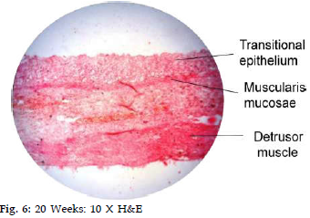
Background: Urinary bladder serves an important function of temporary storage of urine without changing its constituents. Many studies are available for the gastrointestinal tract [1,2,3] ellustrating the muscularis mucosae presence and its role in local contractility. But only few studies are available to describe the development and role of muscularis mucosae layer of urinary bladder. Current study is on the urinary bladder involving the microscopic examination of wall of 50 aborted human fetuses’ urinary bladders of different gestational age from 9th week onwards. Aims: 1. To note presence, structural differentiation and maturity of muscularis mucosae layer which it attains at different stages of development to show the adult picture. 2. To compare and contrast differences between different age groups and with previous studies and available literature. Study Design: Observational (Qualitative) study. Material and Method: 50 aborted human fetuses (29 females and 21 males) of different gestational age from 9th week onwards were collected, urinary bladder were taken out and fixed in a fixative. Blocks of tissues were made from bladder wall proper, trigone & bladder neck and processed to get sections which were stained with 1) Haematoxylin and Eosin [4], 2) Masson’s trichrome stain [4]. Statistical Analysis: No measurements have been taken as it is an Observational study, so statistical analysis is not applicable. Results: The stained sections were examined under light microscope at 10 X and 40 X magnifications. Muscularis mucosa was not differentiable between 12-16 weeks. At 17th week, it was first seen as a thin, discontinuous layer of spindle shaped cells. 24th week onwards muscularis mucosae was thicker, wavy and stained dark eosinophilic. Further it showed adult picture and stained bright red with special muscle stain Masson’s Trichrome. Conclusions: Muscularis mucosa was discernible from 17th week and increased in thickness and differentiated in subsequent weeks. It was found in 75% of studied aborted fetuses.
References
No records found.
About this article

Cite this article
Licence:
| Received | Accepted | Published |
|---|---|---|
| N/A | N/A | N/A |
DOI: http://dx.doi.org/10.21088/ija.2320.0022.6317.17
Keywords
Histogenesis; Muscularis Mucosae; Fetus; Urinary Bladder.ÂSearch for Similar Articles
Similar Articles
- Supernumerary Pulmonary Vein: A Rare Anatomical Variation Observed During Cadave...
- Elongated Styloid Process: Anatomical Variations and Their Clinical Relevance: A...
- Bridging the Gap Between Dissection and Surgery: The Role of Soft Embalming in M...
- A Cadaveric Study of Retro Hepatic Segment of Inferior Vena Cava and Ostia Venae...
- Estimation of Stature from Forearm Length in Medical Undergraduate Students of a...
Article Level Metrics
Last UpdatedTuesday 22 September 2026, 02:42:42 (IST)
5059
Accesses
13
655
00
Citations
NA
NA
NA
Download citation
Article Keywords
Keyword Highlighting
Highlight selected keywords in the article text.
Timeline
| Received | N/A |
| Accepted | N/A |
| Published | N/A |