Full Text (PDF)
Indian Journal of Forensic Medicine and Pathology 13(1):p 53-56, January-March 2020. | DOI: https://doi.org/10.21088/ijfmp.0974.3383.13120.7
Original Article
Granulomatous Mastitis — Clinicopathological Review of 38 Cases: A 3 Year Study with a Brief Review of Literature
Swati Sharma, Manna Valiathan, Riti Bhattacharya, Shankar M Bakkannavar
Author Information
Licence:
Attribution-Non-commercial 4.0 International (CC BY-NC 4.0)This license enables reusers to distribute, remix, adapt, and build upon the material in any medium or format for noncommercial purposes only, and only so long as attribution is given to the creator.
Indian Journal of Forensic Medicine and Pathology 13(1):p 53-56, January-March 2020. | DOI: https://doi.org/10.21088/ijfmp.0974.3383.13120.7
How Cite This Article:
Valiathan M, Sharma S, Bhattacharya R, et al. Granulomatous Mastitis — Clinicopathological Review of 38 Cases: A 3 Year Study with a Brief Review of Literature. Indian J Forensic Med Pathol. 2020;13(1):53–56.Timeline
Received : August 21, 2019
Accepted : November 13, 2019
Published : December 30, 2019
Abstract
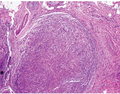
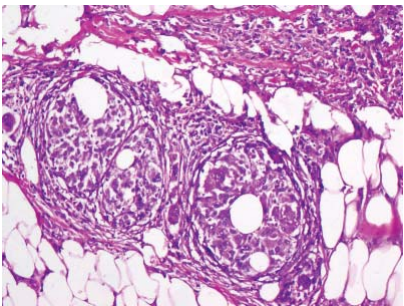
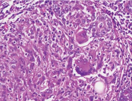
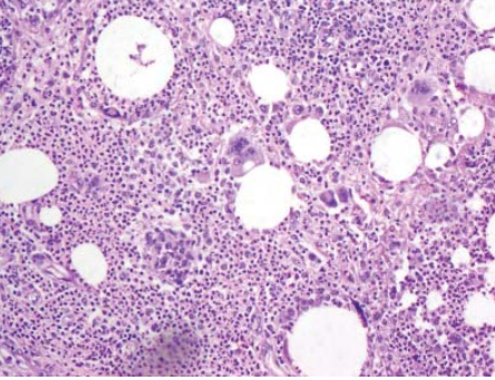
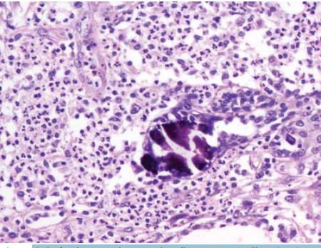
Introduction: Granulomatous mastitis (GM) is a rare, chronic inflammatory condition of obscure etiology, varied clinical and pathological features. Methods: This was a restrospective, three-year analysis, of the clinical and histological parameters of diagnosed cases of GM. Results: An analysis of age at presentation revealed the mean age as 35.3 yr. Clinical data was available for 26 patients and the mean lesional size was 5.3 x 4.3 cm. The most common presenting complaint was as a lump, with diffuse swelling and nipple discharge next in frequency. Clinical diagnosis varied widely from benign to malignant. Treatment administered included incision and curettage, lumpectomy and simple mastectomy. Thirty-eight cases reviewed showed granulomas in all the cases. Caseous necrosis was absent. Special stains for tubercular bacilli and fungi had been done in 26 cases and were negative. Follow-up ranged from 3–5.5 years. Recurrence was documented in 57.6% of patients. Conclusion: GM, generally, is a disease of young women that is of particular significance since it can easily be mistaken for malignancy. Histopathological diagnosis is confirmatory.
References
- 1. Kessler E, Wooloch Y. Granulomatous mastitis: a lesion clinically simulating carcinoma. Am J Clin Pathol. 1972 Dec;58(6):642–6.
- 2. Cohen C. Granulomatous mastitis: a review of 5 cases. S Afr Med J. 1977 Jul 2;52(1):15–6.
- 3. Bhansali M. Granulomatous mastitis: A rare presentation. Bombay Hospital Journal. 2008;50(3):490–92.
- 4. Tuli R, J O’Hara B, Hines J, et al. Idiopathic granulomatous mastitis masquerading as carcinoma of the breast: a case report and review of the literature. Int Semin Surg Oncol. 2007 Jul 27;4:21.
- 5. Larsen LJH, Peyvandi B, Grant E, et al. Granulomatous lobular mastitis: Imaging, Diagnosis, and Treatment. Am J Roentgenol. 2009 Aug;193(2):574–81.
- 6. Ozturk E, Akin M, Can MF, et al. Idiopathic granulomatous mastitis. Saudi Med J. 2009;30(1):45–9.
- 7. Belaabidia B, Essadki O, el Mansouri A, et al. Idiopathic granulomatous mastitis: apropos of 8 cases and review of the literature. Gynecol Obstet Fertil. 2002;30(5):383–9.
- 8. Lai EC, Chan WC, Ma TK, et al. The role of conservative treatment in idiopathic granulomatous mastitis. Breast J. 2005;11:454–6.
- 9. Kocaoglu M, Somuncu I, Ors F, et al. Imaging findings in idiopathic granulomatous mastitis. A review with emphasis on magnetic resonance imaging. J Comput Assist Tomogr. 2004;28:635–41.
- 10. Olsen ML, Dilaveri CA. Idiopathic granulomatous mastitis: a case report of breast abscess. BMJ Case Reports. 2011;10.
- 11. Aghajanzadeh M, Hassanzadeh R, Sefat SA. Granulomatous mastitis: Presentations, diagnosis, treatment and outcome in 206 patients from the north of Iran. Breast. 2015;24(4):456–60.
- 12. Helal TEA, Shash LS, Saad El-Din SA, et al. Idiopathic granulomatous mastitis: cytologic and histologic study of 65 Egyptian patients. Acta Cytologica. 2016;60(5):438–44.
- 13. Deng JQ, Yu L, Yang Y. Steroids administered after vacuum-assisted biopsy in the management of idiopathic granulomatous Mastitis. J Clin Pathol. 2017;70(10):827–31.
- 14. Halim NA, Uthman I, Rammal R, et al. Idiopathic Granulomatous Mastitis Presenting as a Breast Pseudotumor: Case Reports with Review of the Literature. Case Reports in Rheumatology. 2018;2018:5.
- 15. Kiyak G, Dumlu EG, Kilinc I, et al. Management of idiopathic granulomatous mastitis: dilemmas in diagnosis and treatment. BMC Surg. 2014 Sep 4;14:66.
Data Sharing Statement
There are no additional data available. All raw data and code are available upon request.
Funding
This research received no funding.
Author Contributions
Whether all authors contributed significantly to the work and approve its publication.
Ethics Declaration
This article does not involve any human or animal subjects, and therefore does not require ethics approval.
Acknowledgements
We would like to express our gratitude to the patients, their families, and all those who have contributed to this study.
Conflicts of Interest
The authors report no conflicts of interest in this work.
About this article

Cite this article
Valiathan M, Sharma S, Bhattacharya R, et al. Granulomatous Mastitis — Clinicopathological Review of 38 Cases: A 3 Year Study with a Brief Review of Literature. Indian J Forensic Med Pathol. 2020;13(1):53–56.
Licence:
Attribution-Non-commercial 4.0 International (CC BY-NC 4.0)This license enables reusers to distribute, remix, adapt, and build upon the material in any medium or format for noncommercial purposes only, and only so long as attribution is given to the creator.
| Received | Accepted | Published |
|---|---|---|
| August 21, 2019 | November 13, 2019 | December 30, 2019 |
DOI: https://doi.org/10.21088/ijfmp.0974.3383.13120.7
Keywords
CorticosteroidsGranulomaLobulocentricMastitisNecrosisSearch for Similar Articles
Similar Articles
- Diatom Analysis in Thermophilic water bodies: A Study on Hot Water Springs of Ma...
- Morphometric Study of Supra-meatal Triangle and Mastoid process in the Adult dry...
- Histopathological Spectrum of Autopsy Cases: A One Year Retrospective Observatio...
- Forensic Differentiation of Manner of Death in Railway Fatalities: A FourCase Se...
- Understanding assault Patterns: A Cross-sectional study of Cases at Chri, Kelamb...
Article Level Metrics
Last UpdatedSunday 18 October 2026, 08:52:58 (IST)
16253
Accesses
15
3204
00
Citations
NA
NA
NA
Download citation
Article Keywords
Keyword Highlighting
Highlight selected keywords in the article text.
Timeline
| Received | August 21, 2019 |
| Accepted | November 13, 2019 |
| Published | December 30, 2019 |
licence
Attribution-Non-commercial 4.0 International (CC BY-NC 4.0)
This license enables reusers to distribute, remix, adapt, and build upon the material in any medium or format for noncommercial purposes only, and only so long as attribution is given to the creator.