Full Text (PDF)
Indian Journal of Pathology: Research and Practice 14(2):p 68-70, may-august 2025. | DOI: https://doi.org/10.21088/ijprp.2278.148X.14225.3
Case Report
Fine Needle Aspiration Cytology for Diagnosis of Malignant Acinic Cell Carcinoma of the Parotid Gland: A Case Report
Author Information
Licence:
Attribution-Non-commercial 4.0 International (CC BY-NC 4.0)This license
enables reusers to distribute, remix, adapt, and build upon the material in any
medium or format for noncommercial purposes only, and only so long as
attribution is given to the creator.
Indian Journal of Pathology: Research and Practice 14(2):p 68-70, may-august 2025. | DOI: https://doi.org/10.21088/ijprp.2278.148X.14225.3
How Cite This Article:
Devajit Nath, Adity Sarma. Fine Needle Aspiration Cytology for Diagnosis of Malignant Acinic Cell Carcinoma of the Parotid Gland: A Case Report. Ind Jr of Path: Res and Practice 2025; 14(2) 68-70.Timeline
Received : May 14, 2025
Accepted : June 16, 2025
Published : August 30, 2025
Abstract
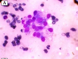
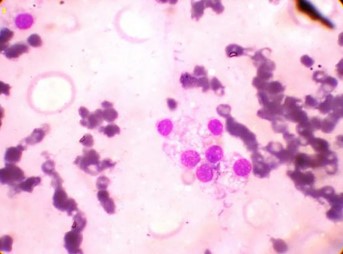
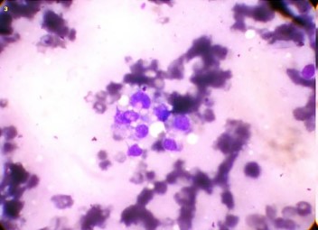
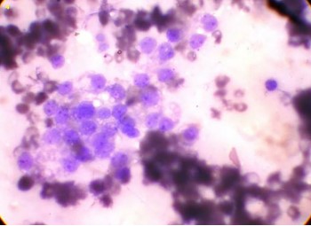
Fine needle aspiration cytology (FNAC) is an essential tool for diagnosing salivary gland tumors. Here we present a case involving a 45-year-old woman with an eight-year history of a slowly growing, painless swelling in her right parotid gland. FNAC showed clusters of polygonal cells with vacuolated cytoplasm, round nuclei, and prominent nucleoli in a bromuscular background, suggestive of acinic cell carcinoma (ACC). Surgical excision and histopathological analysis firm the diagnosis. This case highlights FNAC’s crucial role in the early detection and diagnosis of rare salivary gland malignancies.
References
- 1. Speight P.M., Barrett A.W. Salivary gland tumours. Oral Dis. 2002; 8(5): 229-40.
- 2. Ellis G.L., Auclair P.L. Tumors of the Salivary Glands. Washington: AFIP Atlas of Tumor Pathology; 2008.
- 3. Hoffman H.T., Karnell L.H., Robinson R.A., Pinkston J.A., Menck H.R. National Cancer Data Base report on cancer of the head and neck: Acinic cell carcinoma. Head Neck. 1999; 21(4): 297-309.
- 4. Klijanienko J., Vielh P. Fine-needle sampling of salivary gland lesions III: cytology and histology correlation of 75 cases of acinic cell carcinoma. DiagnCytopathol. 1997; 17(5): 347-52.
- 5. Nagel H., Hotze H.J., Laskawi R., Chilla R., Droese M. Cytologic diagnosis of acinic-cell carcinoma of salivary glands. DiagnCytopathol. 1999; 20(6): 402-12.
- 6. Schindler S., Nayar R., Dutra J., Bedrossian C.W. Diagnostic challenges in aspiration cytology of the salivary glands. Semin Diagn Pathol. 2001; 18(2): 124-46.
- 7. Elsheikh T.M., Bernacki E.G. Fine needle aspiration cytology of cellular pleomorphic adenoma with emphasis on diagnostic pitfalls. DiagnCytopathol. 1996; 14(1): 59-64.
- 8. Seethala R.R., Barnes E.L. Rare malignant tumors of the salivary glands. SurgPathol Clin. 2011; 4(4): 1167-78.
- 9. Skalova A., Michal M., Simpson R.H., et al. Acinic cell carcinoma of salivary glands with high-grade transformation: clinicopathologic study of 9 cases. Am J SurgPathol. 2009; 33(8): 1137-45.
- 10. Vander Poorten V., Triantafyllou A, Thompson L.D., et al. Salivary acinic cell carcinoma: Reappraisal and update. Eur Arch Otorhinolaryngol. 2016; 273(11): 3511-31.
Data Sharing Statement
There are no additional data available. All raw data and code are available upon request.
Funding
This research received no funding.
Author Contributions
All authors contributed significantly to the work and approve its publication.
Ethics Declaration
This article does not involve any human or animal subjects, and therefore does not require ethics approval.
Acknowledgements
We would like to express our gratitude to the patients, their families, and all those who have contributed to this study.
Conflicts of Interest
No conflicts of interest in this work.
About this article

Cite this article
Devajit Nath, Adity Sarma. Fine Needle Aspiration Cytology for Diagnosis of Malignant Acinic Cell Carcinoma of the Parotid Gland: A Case Report. Ind Jr of Path: Res and Practice 2025; 14(2) 68-70.
Licence:
Attribution-Non-commercial 4.0 International (CC BY-NC 4.0)This license
enables reusers to distribute, remix, adapt, and build upon the material in any
medium or format for noncommercial purposes only, and only so long as
attribution is given to the creator.
| Received | Accepted | Published |
|---|---|---|
| May 14, 2025 | June 16, 2025 | August 30, 2025 |
DOI: https://doi.org/10.21088/ijprp.2278.148X.14225.3
Keywords
FNACAcinic cell carcinomaHistopathologyParotid glandSearch for Similar Articles
Similar Articles
- Clinico-Pathological and Immunohistochemical Study of Primary CNS Tumors in a Te...
- The Role of SGLT2 Inhibitors in Cardiovascular Protection Beyond Diabetes: A Co...
- Epidermal Inclusion Cyst Presenting as 4th Ventricle Space Occupying Lesion: A R...
- Silent Retroperitoneal Seminoma Arising from an Occult Undescended Testis: Diagn...
- Scapular Metastasis of Oral Squamous Cell Carcinoma: A Case Report of an Unusual...
Article Level Metrics
Last UpdatedSaturday 17 October 2026, 10:31:18 (IST)
4177
Accesses
16
671
00
Citations
NA
NA
NA
Download citation
Article Keywords
Keyword Highlighting
Highlight selected keywords in the article text.
Timeline
| Received | May 14, 2025 |
| Accepted | June 16, 2025 |
| Published | August 30, 2025 |
licence
Attribution-Non-commercial 4.0 International (CC BY-NC 4.0)
This license
enables reusers to distribute, remix, adapt, and build upon the material in any
medium or format for noncommercial purposes only, and only so long as
attribution is given to the creator.