Full Text (PDF)
Indian Journal of Pathology: Research and Practice 14(2):p 72-79, may-august 2025. | DOI: https://doi.org/10.21088/ijprp.2278.148X.14225.4
Case Report
Beyond the Gross: Unveiling the Microscopic Truth in 3 Autopsy Cases
Manoj B. Patekar, Abhishek Bhimrao Muley, Ketaki Jayant Patil, Nikhil S. Jagtap
Author Information
Licence:
Attribution-Non-commercial 4.0 International (CC BY-NC 4.0)This license
enables reusers to distribute, remix, adapt, and build upon the material in any
medium or format for noncommercial purposes only, and only so long as
attribution is given to the creator.
Indian Journal of Pathology: Research and Practice 14(2):p 72-79, may-august 2025. | DOI: https://doi.org/10.21088/ijprp.2278.148X.14225.4
How Cite This Article:
Abhishek Bhimrao Muley, Manoj B. Patekar, et al. Beyond the Gross: Unveiling the Microscopic Truth in 3 Autopsy Cases. Ind Jr of Path: Res and Practice 2025; 14(2) 72-79.Timeline
Received : May 08, 2025
Accepted : June 06, 2025
Published : August 30, 2025
Abstract
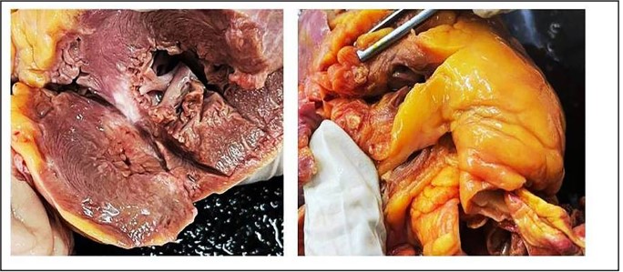
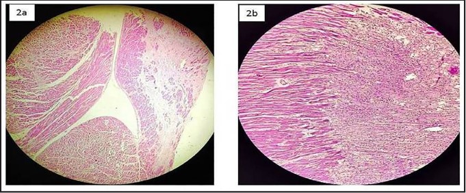
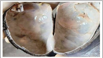
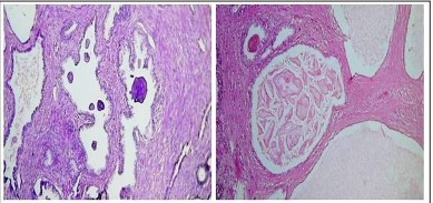
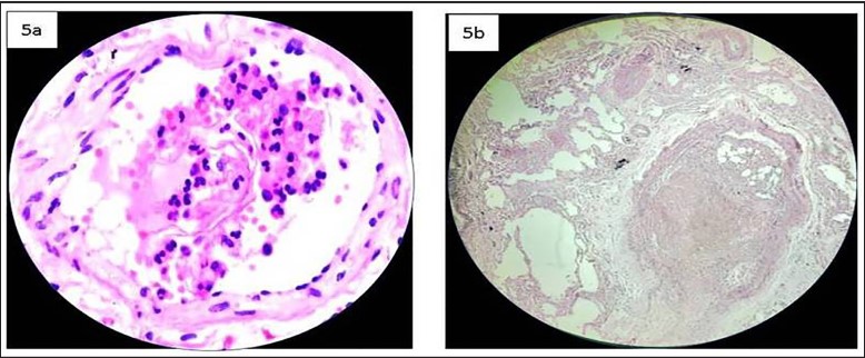
Histopathology, the microscopic examination of tissues, is a keystone of the autopsy examination, providing critical understandings outside what can be observed with the gross examination. In medicolegal autopsies, where the cause and manner of death may be unclear or contested, histopathology plays an especially vital role. This case series emphasizes the important role of histopathological examination in medicolegal autopsies. We present three cases where microscopic tissue exploration through histopathology provided essential information that significantly impacted the final determination of the cause and/ or manner of death. In the first case, histopathology revealed subtle myocardial changes indicative of a previously unsuspected cardiac event. The second case demonstrated the utility of histopathology in distinguishing death due to rupture of hepatic cyst from alleged death due to blunt trauma. Finally, the third case highlights how microscopic evaluation identified a lethal but grossly undetectable disease process related to amniotic fluid embolism in a postoperative case. These cases illustrate that histopathology is a crucial tool in medicolegal investigations, often providing critical evidence that macroscopic gross examination alone cannot.
References
- 1. Shubhendu K., Mundri S., Kumar S., Kumar A. The Impact of Histopathology on Medical Board Autopsies. Cureus. 2025 Feb 7; 17(2): e78675. doi: 10.7759/cureus.78675. PMID:40062106; PMCID: PMC11890611.
- 2. Autopsy [Internet]. Wikipedia; [cited 2025 Apr 04]. Available from: https://en.wikipedia.org/wiki/Autopsy.
- 3. Histo-Pathology Examination in Medico-legal Autopsy Pros & Cons. J Indian Acad Forensic Med, 32(2).128-131.
- 4. Rao N.G. Practical Forensic Medicine. 3rd Edition, 2007.149.
- 5. Molina D.K., Wood L.E., Frost R.F. Is routine histopathological examination beneficial in all medicolegal autopsies? Am J Forensic Med Pathol. 2007; 28: 1-3.
- 6. Guareschi E.E. The Role of Histology in Forensic Investigations. Preprints.org (www.preprints. org) | Not Peer-Reviewed | Posted: 20 March 2024 doi:10.20944/preprints202403.1133.v1
- 7. Mukherjee B., Ropmay, Slong D., et al. (March 23, 2025) Role of Histopathology in Determining the Cause of Death in Medicolegal Autopsies. Cureus 17(3): e81055. doi:10.7759/cureus.81055
- 8. Banwari M. An erroneous opinion on a cause of death in a forensic autopsy: a case report. Afr Health Sci. 2017 Dec; 17(4): 1246-1249. doi: 10.4314/ahs.v17i4.36. PMID: 29937899; PMCID: PMC5870277.
- 9. Jhajj K.K., Nibhoria S. et al. A study of histopathological examination in medicolegal Autopsies in Faridkot, Punjab.Ind.Journal of FMT: 2013. 7(1): 76-81.
- 10. Zenati H. Hydatid peritonitis caused by liver hydatid cyst rupture into the peritoneal cavity: A case report Hanen Zenati. International Journal of Surgery Case Reports
- 11. Noone P.H. Training in Forensic Histopathology-Significance in Postgraduate Curriculum of Forensic Medicine. Journal of Evidence Based Medicine and Health Care. September 2014. 1(7): 595-598.
- 12. McDougall, Robert J and Graeme J Duke. “Amniotic Fluid Embolism Syndrome: Case Report and Review.” Anaesthesia and Intensive Care 23.1995: 735-740.
- 13. Schandl C.A., Collins K.A. Maternal Autopsy. In: Collins K.A. editor. Autopsy performance and reporting. 2nd ed. Northfield (IL): College of American Pathologists; 2002. p. 135-49.
- 14. Hikiji W., Tamura N., Shigeta A., et al. Fatal amniotic fluid embolism with typical pathohistological, histochemical and clinical features. Forensic Sci Int. 2013 Mar 10; 226 (1-3): e16-9. PMID: 23273942. https://doi. org/10.1016/j.forsciint.2012.12.008.
- 15. Role of Histopathological Examination in Medicolegal Autopsies in Unravelling Precise Causes of Mortality. National Journal of Laboratory Medicine. 2021 Oct, Vol-10(4): PO01-PO04.
Data Sharing Statement
There are no additional data available. All raw data and code are available upon request.
Funding
This research received no funding.
Author Contributions
All authors contributed significantly to the work and approve its publication.
Ethics Declaration
This article does not involve any human or animal subjects, and therefore does not require ethics approval.
Acknowledgements
We would like to express our gratitude to the patients, their families, and all those who have contributed to this study.
Conflicts of Interest
No conflicts of interest in this work.
About this article

Cite this article
Abhishek Bhimrao Muley, Manoj B. Patekar, et al. Beyond the Gross: Unveiling the Microscopic Truth in 3 Autopsy Cases. Ind Jr of Path: Res and Practice 2025; 14(2) 72-79.
Licence:
Attribution-Non-commercial 4.0 International (CC BY-NC 4.0)This license
enables reusers to distribute, remix, adapt, and build upon the material in any
medium or format for noncommercial purposes only, and only so long as
attribution is given to the creator.
| Received | Accepted | Published |
|---|---|---|
| May 08, 2025 | June 06, 2025 | August 30, 2025 |
DOI: https://doi.org/10.21088/ijprp.2278.148X.14225.4
Keywords
HistopathologyAllegationEmbolismHydatid cystManner of deathSearch for Similar Articles
Similar Articles
- Clinico-Pathological and Immunohistochemical Study of Primary CNS Tumors in a Te...
- The Role of SGLT2 Inhibitors in Cardiovascular Protection Beyond Diabetes: A Co...
- Epidermal Inclusion Cyst Presenting as 4th Ventricle Space Occupying Lesion: A R...
- Silent Retroperitoneal Seminoma Arising from an Occult Undescended Testis: Diagn...
- Scapular Metastasis of Oral Squamous Cell Carcinoma: A Case Report of an Unusual...
Article Level Metrics
Last UpdatedSaturday 17 October 2026, 10:26:54 (IST)
4177
Accesses
13
671
00
Citations
NA
NA
NA
Download citation
Article Keywords
Keyword Highlighting
Highlight selected keywords in the article text.
Timeline
| Received | May 08, 2025 |
| Accepted | June 06, 2025 |
| Published | August 30, 2025 |
licence
Attribution-Non-commercial 4.0 International (CC BY-NC 4.0)
This license
enables reusers to distribute, remix, adapt, and build upon the material in any
medium or format for noncommercial purposes only, and only so long as
attribution is given to the creator.