Full Text (PDF)
Indian Journal of Pathology: Research and Practice 13(1):p 13-17, January - March 2024. | DOI: https://doi.org/10.21088/ijprp.2278.148X.13124.2
Original Article
Demographic Profile of CNS Tumors and Histopathological Evaluation of Pituitary Lesions: An Institutional Study
Author Information
Licence:
Attribution-Non-commercial 4.0 International (CC BY-NC 4.0)This license enables reusers to distribute, remix, adapt, and build upon the material in any medium or format for noncommercial purposes only, and only so long as attribution is given to the creator.
Indian Journal of Pathology: Research and Practice 13(1):p 13-17, January - March 2024. | DOI: https://doi.org/10.21088/ijprp.2278.148X.13124.2
How Cite This Article:
Jayasree Geothe, Lekshmy K S. Demographic Profile of CNS Tumors and Histopathological Evaluation of Pituitary Lesions: An Institutional Study. Ind Jr of Path: Res and Practice 2024;13(1) 13-17.Timeline
Received : January 26, 2023
Accepted : March 03, 2023
Published : March 30, 2023
Abstract
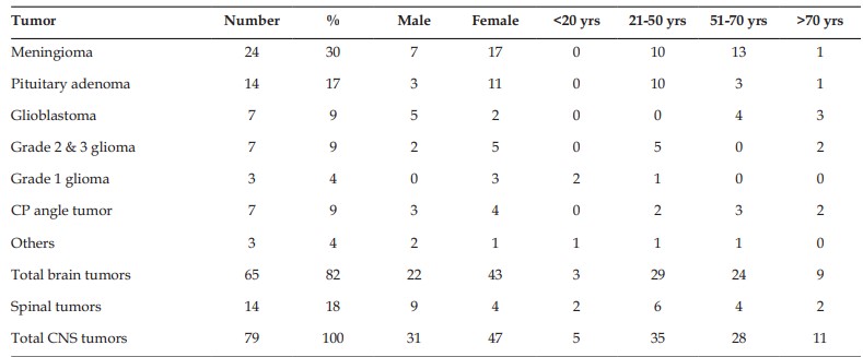
Primary Central Nervous System tumors (CNS trs) are a diverse group of neoplasm showing significant geographic variations in incidence, morbidity, mortality, and survival. Pituitary adenomas (PA) recently termed as Pituitary Neuroendocrine Tumours (PitNET) are the commonest mass in the sellar region, can produce local, and distant effects.1 However, some developmental, inflammatory, and neoplastic lesions may primarily or secondarily involve the sella, mimicking pituitary tumors; recognition of which is crucial for the appropriate management. Aims: To evaluate the histopathological profile of primary CNS trs and to correlate with age, sex and site at presentation. Pituitary lesions are further analyzed to find out prevalence of non adenomatous lesions in pituitary Materials and methods: Histologically diagnosed 79 primary CNS trs and 18 pituitary lesions are analyzed. Results:Meningioma is the commonest brain and CNS trs. The PA being the second commonest CNS trs, constitute 17% of CNS trs, 22% of brain tumors and 78% of pituitary lesions. Other sellar lesions were single cases of Rathkeys cleft cyst (RCC), Arachnoid cyst, craniopharyngioma and pituitary apoplexy. Conclusion: This retrospective study established a baseline profile of brain tumors and sellar lesions based primarily on the histopathological experience at a tertiary care hospital in the Southern Kerala.
References
- 1. WHO Classification of Tumours Endocrine and Neuroendocrine tumours, vol. 8. 5th edn. (International Agency for Research on Cancer, Lyon, France, 2022) https://tumourclassification.iarc.who.int
- 2. Ezzat S, Asa SL, Couldwell WT, Barr CE, Dodge WE, Vance ML, et al. The prevalence of pituitary adenomas: a systematic review. Cancer. 2004;101(3):613-9.
- 3. USG guided Trucut biopsy from left breast lesion Karmarkar VS, Deopujari CE. Sellar Pathologies Mimicking Pituitary Tumors. Neurol India 2020;68, Suppl S1:154-60
- 4. Histopathological profile of brain tumors: A 12- year retrospective study from Madinah, Saudi Arabia,Mohammed Albasri Abdulkader, Hamdan Almuhamdi Nawal, Homoud Alqaidi Sara:Asian Journal of Neurosurgery 2019; 14 ( 4 ) 1106-1111.
- 5. Saeger W, Lüdecke DK, Buchfelder M, Fahlbusch R, Quabbe HJ, Petersenn S. Pathohistological classification of pituitary tumors: 10 years of experience with the German Pituitary Tumor Registry. Eur J Endocrinol. 2007 Feb;156(2):203-16. doi: 10.1530/eje.1.02326. PMID: 17287410.
- 6. Shimon I, Benbassat C, Hadani M. Effectiveness of long-term cabergoline treatment for giant prolactinoma: study of 12 men. Eur J Endocrinol. 2007 Feb;156(2):225-31. doi:10.1530/EJE-06-0646.PMID: 17287412.
- 7. Dekkers, O.M., Karavitaki, N. & Pereira, A.M. The epidemiology of aggressive pituitary tumors (and its challenges). Rev Endocr Metab Disord –209 ,21 2020) 212). https://doi.org/10.1007/s11154-020-09556-7
- 8. Dhandapani S, Singh H, Negm HM, Cohen S, Anand VK, Schwartz TH. Cavernous Sinus Invasion in Pituitary Adenomas: Systematic Review and Pooled Data Meta-Analysis of Radiologic Criteria and Comparison of Endoscopic and Microscopic Surgery. World Neurosurg. 2016 Dec;96:36-46. doi: 10.1016/j.wneu.2016.08.088. Epub 2016 Aug 30.
- 9. de Almeida Verdolin A, Lamback EB, Ventura N, Guasti A, da Mata Pereira PJ, Gadelha MR, Chimelli L. Collision sellar lesions: coexistence of pituitary adenoma and Rathke cleft cyst-a single-center experience. Endocrine. 2020 Apr;68(1):174-181. doi: 10.1007/s12020-019-02149
- 10. Epub 2019 Dec 4. PMID: 31802354.
- 11. Sumida M, Migita K, Tominaga A, Iida K, Kurisu K. Concomitant pituitary adenoma and Rathke’s cleft cyst. Neuroradiology. 2001 Sep;43(9):755-9. doi: 10.1007/s002340100559. PMID: 11594426.
- 12. Gao M, An Y, Huang Z, Niu J, Yuan X, Bai Y, Guo L. The Coexistence of Rathke Cleft Cyst and Pituitary Adenoma. J Craniofac Surg. 2016 Mar;27(2):e128-30. doi:10.1097/SCS.0000000000002371. PMID: 26845092.
- 13. Biousse, V et al. “Precipitating factors in pituitary apoplexy.” Journal of neurology, neurosurgery, and psychiatry vol. 71,4 (2001): 542-5. doi:10.1136/jnnp.71.4.542
- 14. Ranabir, Salam, and Manash P Baruah. “Pituitary apoplexy.” Indian journal of endocrinology and metabolism vol. 15 Suppl 3,Suppl3 (2011): S188-96. doi:10.4103/2230-8210.84862
Data Sharing Statement
There are no additional data available. All raw data and code are available upon request.
Funding
This research received no funding.
Author Contributions
All authors contributed significantly to the work and approve its publication.
Ethics Declaration
This article does not involve any human or animal subjects, and therefore does not require ethics approval.
Acknowledgements
We would like to express our gratitude to the patients, their families, and all those who have contributed to this study.
Conflicts of Interest
No conflicts of interest in this work.
About this article

Cite this article
Jayasree Geothe, Lekshmy K S. Demographic Profile of CNS Tumors and Histopathological Evaluation of Pituitary Lesions: An Institutional Study. Ind Jr of Path: Res and Practice 2024;13(1) 13-17.
Licence:
Attribution-Non-commercial 4.0 International (CC BY-NC 4.0)This license enables reusers to distribute, remix, adapt, and build upon the material in any medium or format for noncommercial purposes only, and only so long as attribution is given to the creator.
| Received | Accepted | Published |
|---|---|---|
| January 26, 2023 | March 03, 2023 | March 30, 2023 |
DOI: https://doi.org/10.21088/ijprp.2278.148X.13124.2
Keywords
Pituitary AdenomasPituitary Neuroendocrine Tumours (Pitnets) MacroadenomasRathkey Cleft CystPituitary ApoplexyCNS TumorsSearch for Similar Articles
Similar Articles
- Clinico-Pathological and Immunohistochemical Study of Primary CNS Tumors in a Te...
- The Role of SGLT2 Inhibitors in Cardiovascular Protection Beyond Diabetes: A Co...
- Epidermal Inclusion Cyst Presenting as 4th Ventricle Space Occupying Lesion: A R...
- Silent Retroperitoneal Seminoma Arising from an Occult Undescended Testis: Diagn...
- Scapular Metastasis of Oral Squamous Cell Carcinoma: A Case Report of an Unusual...
Article Level Metrics
Last UpdatedSunday 20 September 2026, 22:02:29 (IST)
3662
Accesses
11
541
00
Citations
NA
NA
NA
Download citation
Article Keywords
Keyword Highlighting
Highlight selected keywords in the article text.
Timeline
| Received | January 26, 2023 |
| Accepted | March 03, 2023 |
| Published | March 30, 2023 |
licence
Attribution-Non-commercial 4.0 International (CC BY-NC 4.0)
This license enables reusers to distribute, remix, adapt, and build upon the material in any medium or format for noncommercial purposes only, and only so long as attribution is given to the creator.