Full Text (PDF)
Indian Journal of Pathology: Research and Practice 15(2):p 163-165, May - Aug 2026. | DOI: https://doi.org/10.21088/ijprp.2278.148X.15226.10
Case Report
Epidermal Inclusion Cyst Presenting as 4th Ventricle Space Occupying Lesion: A Rare
Syeda Iqra Usman, Mohammad Feroz Alam, Bushra Siddiqui
Author Information
Licence:
Attribution-Non-commercial 4.0 International (CC BY-NC 4.0)This license enables reusers to distribute, remix, adapt, and build upon the material in any medium or format for noncommercial purposes only, and only so long as attribution is given to the creator.
Indian Journal of Pathology: Research and Practice 15(2):p 163-165, May - Aug 2026. | DOI: https://doi.org/10.21088/ijprp.2278.148X.15226.10
How Cite This Article:
Syeda Iqra Usman, Mohammad Feroz Alam, Bushra Siddiqui. Epidermal Inclusion Cyst Presenting as 4th Ventricle Space Occupying Lesion: A Rare. Ind Jr of Path: Res and Practice 2026; 15(2) 163-165.Timeline
Received : December 16, 2025
Accepted : January 18, 2026
Published : August 30, 2026
Abstract
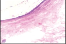
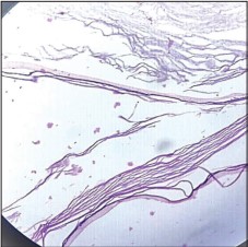
Background: Epidermal inclusion (epidermoid) cysts are rare benign intracranial lesions that account for less than 1% of all intracranial tumors. Their occurrence within the fourth ventricle is exceptionally uncommon and may lead to significant mass-effect symptoms. Case Presentation: A 45-year-old female presented with a one-year history of intermittent headache with recent worsening, followed by progressive vertigo and projectile vomiting. Neuroimaging (CT and MRI) demonstrated a wellcircumscribed cystic lesion occupying the fourth ventricle with extension into the bilateral cerebellar hemispheres and foramina of Luschka and Magendie, producing marked compression of the pons, medulla, and cerebellar hemispheres. The lesion was surgically excised. Histopathological examination revealed a cyst lined by stratified squamous epithelium containing abundant keratin flakes, confirming the diagnosis of an epidermal inclusion cyst. The postoperative period was uneventful, and the patient experienced complete resolution of symptoms. Conclusion: Fourth ventricular epidermal inclusion cysts are rare lesions that may mimic other posterior fossa tumors. Radiological features combined with histopathological evaluation are essential for accurate diagnosis. Surgical excision remains the treatment of choice and is associated with favourable outcomes when critical neurovascular structures are preserved.
References
- 1. Mohan A., Unni C., Kannoth P., Parambil R.M. A Rare Case of Giant Intradiploeic Epidermal Cyst of the Frontal Bone with Intracranial Extension. Asian J Neurosurg. 2020; 15: 670-3.
- 2. Wei Y., Chen P., Wu H. Gigantic occipital epidermal cyst in a 56-year-old female: A case report. World J Clin Cases. 2024; 12: 1169-73.
- 3. Ali A.A., Tahir S.M., Memon A.S., Dahri A.A. Epidermoid inclusion cyst of the perineum: A rare case report in a 50 years old male. J Ayub Med Coll Abbottabad. 2009; 21: 179–80.
- 4. Solanki S.P., Maccormac O., Dow G.R., Smith S. Malignant transformation of residual posterior fossa epidermoid cyst to squamous cell carcinoma. Br J Neurosurg. 2017; 31: 497–8.
- 5. Netsky M.G. Epidermoid tumors. Review of the literature. Surg Neurol. 1988;29:477–83.
- 6. Patibandla M., Yerramneni V., Mudumba V., Manisha N., Addagada G. Brainstem epidermoid cyst: an update. Asian J Neurosurg. 2016; 11: 194–200.
- 7. Pikis S., Cohen J.E., Margolin E. Basilar artery dissection: a rare complication of posterior fossa epidermoid cyst resection, and evaluation of the possible effects of cerebrospinal fluid drainage on disease progression. J Clin Neurosci. 2016; 32: 141–143.
- 8. McLendon R.E., Rosenblum M.K., Bigner D.D. Russell and Rubinstein’s Pathology of Tumors of the Nervous System 7Ed. Hodder Arnold; 2006:583–90.
- 9. Mortini P., Bailo M., Spina A., Acerno S., Boari N., Gagliardi F. Cyst-cisternal shunting for cystic multirecurrent brainstem epidermoid: case report and literature review. Acta Neurochir (Wien). 2016; 158: 1197–1201.
- 10. 10-Akar Z., Tanriover N., Tuzgen S., Kafadar A.M., Kuday C. Surgical treatment of intracranial epidermoid tumors. Neurol Med Chir (Tokyo). 2003; 43: 275–80.
Data Sharing Statement
There are no additional data available. All raw data and code are available upon request.
Funding
This research received no funding.
Author Contributions
All authors contributed significantly to the work and approve its publication.
Ethics Declaration
This article does not involve any human or animal subjects, and therefore does not require ethics approval.
Acknowledgements
We would like to express our gratitude to the patients, their families, and all those who have contributed to this study.
Conflicts of Interest
No conflicts of interest in this work.
About this article

Cite this article
Syeda Iqra Usman, Mohammad Feroz Alam, Bushra Siddiqui. Epidermal Inclusion Cyst Presenting as 4th Ventricle Space Occupying Lesion: A Rare. Ind Jr of Path: Res and Practice 2026; 15(2) 163-165.
Licence:
Attribution-Non-commercial 4.0 International (CC BY-NC 4.0)This license enables reusers to distribute, remix, adapt, and build upon the material in any medium or format for noncommercial purposes only, and only so long as attribution is given to the creator.
| Received | Accepted | Published |
|---|---|---|
| December 16, 2025 | January 18, 2026 | August 30, 2026 |
DOI: https://doi.org/10.21088/ijprp.2278.148X.15226.10
Keywords
Epidermal inclusion cystEpidermoid cystFourth ventriclePosterior fossa lesionIntracranial cystSearch for Similar Articles
Similar Articles
- The Role of SGLT2 Inhibitors in Cardiovascular Protection Beyond Diabetes: A Co...
- Silent Retroperitoneal Seminoma Arising from an Occult Undescended Testis: Diagn...
- Scapular Metastasis of Oral Squamous Cell Carcinoma: A Case Report of an Unusual...
- Adrenocortical Carcinoma Presenting as a Retroperitoneal Mass: Diagnostic Challe...
- Unicentric Castleman Disease of the Neck Mimicking Lymphoma: A Diagnostic Challe...
Article Level Metrics
Last UpdatedMonday 24 August 2026, 12:19:34 (IST)
2915
Accesses
2
472
00
Citations
NA
NA
NA
Download citation
Article Keywords
Keyword Highlighting
Highlight selected keywords in the article text.
Timeline
| Received | December 16, 2025 |
| Accepted | January 18, 2026 |
| Published | August 30, 2026 |
licence
Attribution-Non-commercial 4.0 International (CC BY-NC 4.0)
This license enables reusers to distribute, remix, adapt, and build upon the material in any medium or format for noncommercial purposes only, and only so long as attribution is given to the creator.