Full Text (PDF)
Indian Journal of Anatomy 11(1):p 9-14, January-March 2022. | DOI: https://doi.org/10.21088/ija.2320.0022.11122.1
Original Article
Comparative Study of Foramen Magnum in Dry Cadaveric Skulls and Computerized Tomography Images in North Interior Part of Karnataka Region
Ishwar B Bagoji null, 1Mohammad Tahir K Qureshi null, 2Ishwar B Bagoji 3R S Bulagouda null, 4Gavishiddappa A Hadimani null, 5M Lakshmi Narayan null, 6Anand V Nimbal null
Author Information
Licence:
Indian Journal of Anatomy 11(1):p 9-14, January-March 2022. | DOI: https://doi.org/10.21088/ija.2320.0022.11122.1
How Cite This Article:
M K Qureshi, R S Bulagouda, Gavishiddappa A. Hadimani et al. /Comparative Study of Foramen Magnum in Dry Cadaveric Skulls and Computerized Tomography Images in North Interior Part of Karnataka Region./Indian J Anat.
2022;11(1):9-14
Timeline
Received : January 02, 2022
Accepted : February 12, 2022
Published : March 01, 2022
Abstract
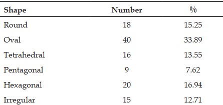
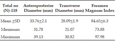
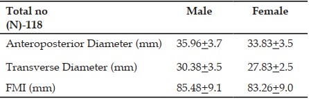
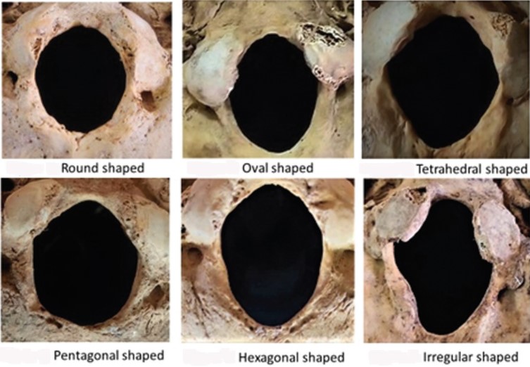
The foramen magnum (FM) is a unique and complex anatomical opening in the base of the skull,which the posterior cranial fossa communicates with the vertebral canal. It is also related to a number of pathological conditions, including Chiari malformations, various tumours, and occipital dysplasias. The study aimed to compare and evaluate the foramen magnummorphology in dry skulls and CT scan images. Material and methods: The morphology of the foramen magnum was assessed in 118 adult non-pathological dry human skulls and 3D computer tomography images in 118 individuals (66 male, 52 female) aged 18–75 years. Result: the incidence of various shapes of the foramen magnum was observed the most common shape was oval (33.89%) in skulls, followed by round (15.25%), tetrahedral (13.55%), pentagonal (7.32%), hexagonal (16.94%) and irregular (12.71%). The mean anteroposterior 33.76 ±2.1mm & 28.09+ 1.9 mm transverse diameter of the foramen magnum. The minimum and maximum values for AP diameter were 31.78 & 39.13. Inthe CT scan study, the mean anteroposterior diameter of FM in the male 35.96± 3.7 and 33.85±3.5 in females. The transverse diameter in males 30.38±3.5 and 27.83±2.5 in females. The FMI was observed in males 85.48±9.1 and 83.26±9.0 in female. Discussion: Results provides baseline data for the anatomist and important information for neurosurgeons to approach the cranial base with maximum safety and minimum mortality and morbidity.
References
No records found.
Data Sharing Statement
There are no additional data available. All raw data and code are available upon request.
Funding
This research received no funding.
Author Contributions
All authors contributed significantly to the work and approve its publication
Ethics Declaration
Provide information related to the Ethics Committee approval with approval number OR write This article does not involve any human or animal subjects, and therefore does not require ethics approval.
Acknowledgements
We would like to express our gratitude to the patients, their families, and all those who have contributed to this study.
Conflicts of Interest
The authors report no conflicts of interest in this work.
About this article

Cite this article
M K Qureshi, R S Bulagouda, Gavishiddappa A. Hadimani et al. /Comparative Study of Foramen Magnum in Dry Cadaveric Skulls and Computerized Tomography Images in North Interior Part of Karnataka Region./Indian J Anat.
2022;11(1):9-14
Licence:
| Received | Accepted | Published |
|---|---|---|
| January 02, 2022 | February 12, 2022 | March 01, 2022 |
DOI: https://doi.org/10.21088/ija.2320.0022.11122.1
Keywords
Foramen magnummorphological variationCervico-medullary junctionTranscondylar approach.morphological variationCervico-medullary junctionTranscondylar approach.Search for Similar Articles
Similar Articles
- Supernumerary Pulmonary Vein: A Rare Anatomical Variation Observed During Cadave...
- Elongated Styloid Process: Anatomical Variations and Their Clinical Relevance: A...
- Bridging the Gap Between Dissection and Surgery: The Role of Soft Embalming in M...
- A Cadaveric Study of Retro Hepatic Segment of Inferior Vena Cava and Ostia Venae...
- Estimation of Stature from Forearm Length in Medical Undergraduate Students of a...
Article Level Metrics
Last UpdatedSunday 20 September 2026, 22:42:17 (IST)
5038
Accesses
17
601
00
Citations
NA
NA
NA
Download citation
Article Keywords
Keyword Highlighting
Highlight selected keywords in the article text.
Timeline
| Received | January 02, 2022 |
| Accepted | February 12, 2022 |
| Published | March 01, 2022 |