Full Text (PDF)
Indian Journal of Forensic Medicine and Pathology 18((2 Suppl)):p 191-203, April-June 2025. | DOI: https://doi.org/10.21088/ijfmp.0974.3383.18225.19
Original Article
Characterization of the Mammalian Post-mortem Thanatomicrobiome: A Time and Temperature Based Approach using Culture Dependent and Independent Techniques
Neeharika Srivastava, Harshita Tiwari, Tripti Bhatnagar
Author Information
Licence:
Attribution-Non-commercial 4.0 International (CC BY-NC 4.0)This license enables reusers to distribute, remix, adapt, and build upon the material in any medium or format for noncommercial purposes only, and only so long as attribution is given to the creator.
Indian Journal of Forensic Medicine and Pathology 18((2 Suppl)):p 191-203, April-June 2025. | DOI: https://doi.org/10.21088/ijfmp.0974.3383.18225.19
How Cite This Article:
Tiwari H, Bhatnagar T, Srivastava N. Characterization of the Mammalian Post-mortem Thanatomicrobiome: A Time and Temperature Based Approach using Culture Dependent and Independent Techniques. Indian J Forensic Med Pathol. 2025;18(2 Suppl):191-203.Timeline
Received : June 25, 2024
Accepted : June 21, 2025
Published : June 30, 2025
Abstract
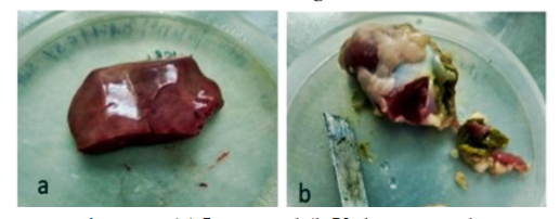
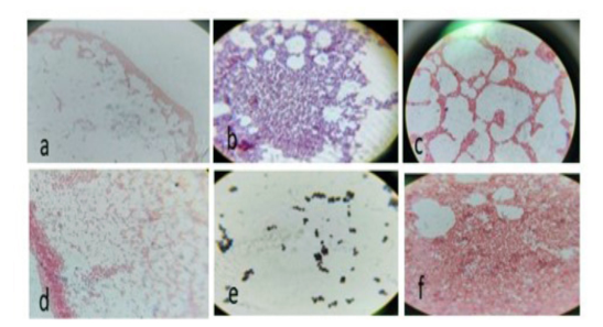
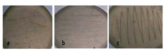
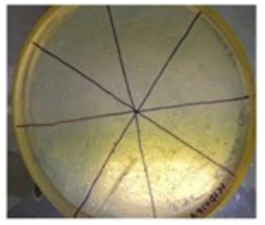
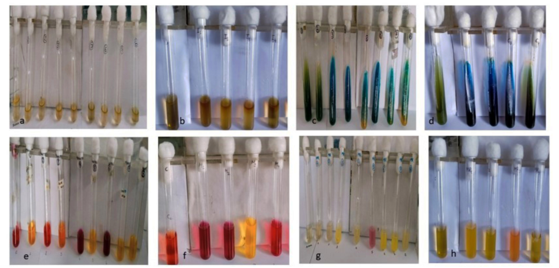
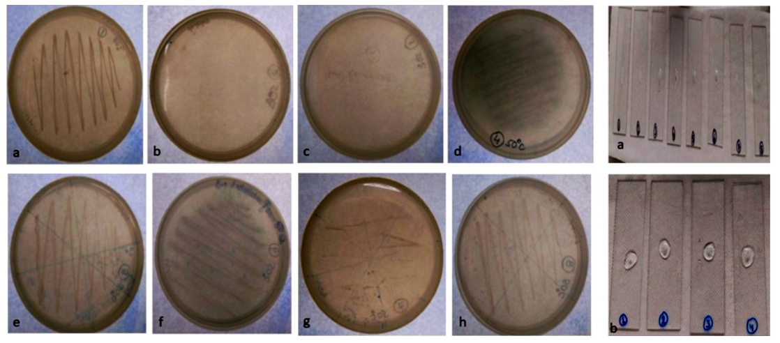
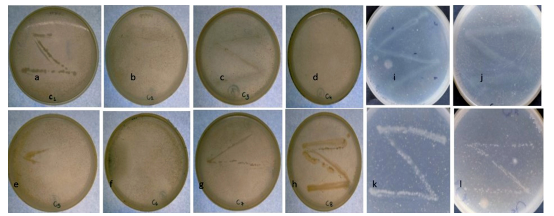
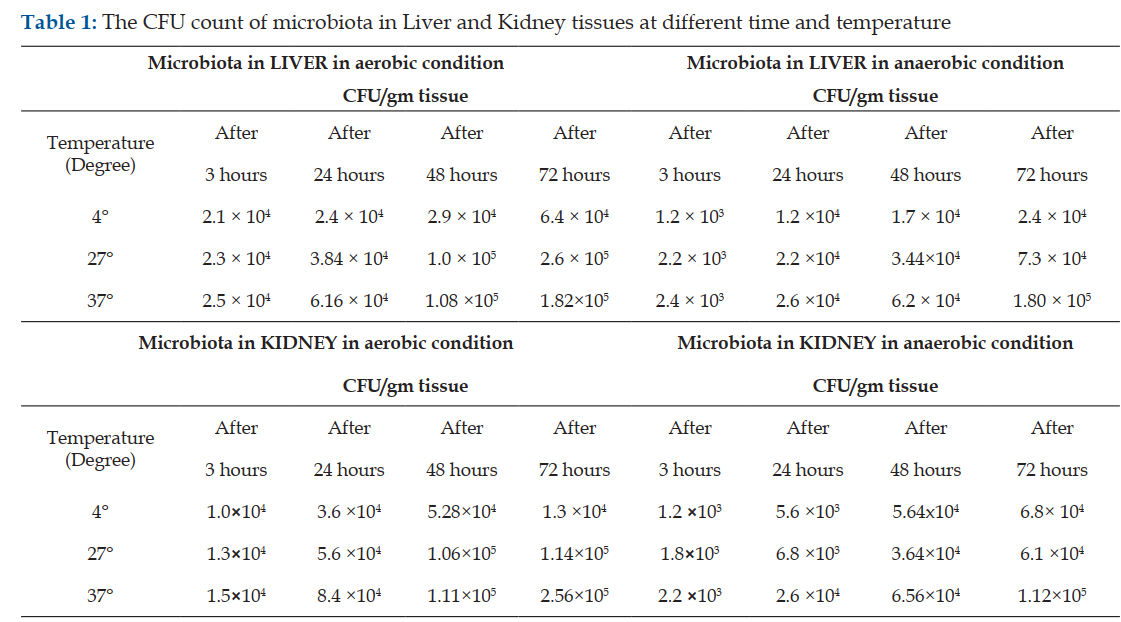
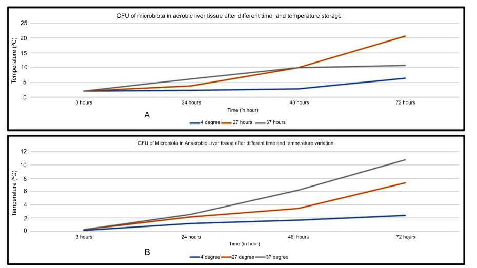
Introduction: The thanatomicrobiome (Thanatos, Greek for death) or the postmortem microbiomes have been recognized as a significant biomarker to determine time and whereabouts of host death. The number and abundance of each organ’s signature microbioata can be of utmost importance to forensic microbiologists as a novel data source for estimating post-mortem interval. Method: Fresh samples of Liver and Kidney of goat were taken and processed. Cultures were prepared and incubated at varying temperatures (37°C, 27°C, and 4°C) in both aerobic and anaerobic condition. After every 24hour, the plates were observed to examine the growth for isolation of microbial colonies. Results: The present finding focuses on the characterization of various thanatomicrobiome compositions across time, temperature and oxygen availability once the animal is passed out. Conclusion: In the present study it was found that increase in time after death leads to variation in bacterial culture in the internal tissues of body which helps in estimation of Post-mortem interval (PMI) in different conditions (such as aerobic and anaerobic), and at different storage temperature.
References
- 1. Alan, G., and Sarah, J.P., Microbes as forensic indicators. Trop Biomed. 29, 311–330, 2012.
- 2. Chauhan A.S., Srivastava, N., Kehri H.K., and Sharma B., “Optimization of culture conditions for some identified fungal species and stability profile of 𝛼-galactosidase produced,” Biotechnology Research International, vol. 2013, Article ID 920759, 8 pages, 2013.
- 3. Hooper, L.V., Gordon, J.I., Commensal host – bacterial relationships in the gut. Science, 292: 1115–1118, 2001.
- 4. Ley R.E., Backhed F., Turnbaugh P., et al., Obesity alters gut microbial ecology, Proc Natl Acad Sci., USA., 102:11070–11075, 2005. DOI: 10.1073/pnas.0504978102
- 5. Hyde, E.R., Harmann, D.P., Lynne, A.M., Bucheli, S.R., and Petrosino, J.F., The bacterial community structure of a corpse at the beginning and end of the bloat stage of decomposition is wknown as “the living dead., 8(10): e77733, 2013. DOI 10.1371/journal.pone.0077733.
- 6. Tuomisto, S., Pessi, T., Collin, P., Vuento, R., Aittoniemi, J., and Karhunen, P.J., Alterations in gut bacterial populations and their translocation in liver and ascites in alcoholic liver cirrhotic, Bmc Gastroenterology, 14:1-8, 2014. Doi: 10.1186/1471-230X-14-40.
- 7. Can, I., Javan, G.T., Pozhitkov, A.E., and Noble, P.A., Unique thanatomicrobiome signatures are detected in human blood and internal organs, Journal of Microbiological, 2014 Doi: 10.1016/j.mimet.2014.07.026 J. Microbial. Methods 106, 1–7.
- 8. Damann, F.E., Williams, D.E., and Layton, A.C., Possibility of measuring post-mortem period via bacterial community succession in deteriorating human bone. J. Forensic Sci. 60, 844–850, 2015. Doi: 10.1111/1556-4029.12744.
- 9. Hauther, K.A., Cobaugh, K.L., Jantz, L.M., Sparer, T.E., and debruyn, J.M., Using postmortem human gug microbial populations to calculate the period since death. J. Forensic Sci., 60: 1234–1240,2015. Doi: 10.1111/1556- 4029.12828.
- 10. Kelsey E. Lawrence, et.al., Effect of temperature and time on the thanatomicrobiome of the cecum, ileum, kidney, and lung of domestic rabbits, 2: 155-163, 2019. Doi/10.1177/1040638719828412.
- 11. Ford, W.W., On the bacteriology of normal organs. J. Hyg. (Lond.), 1: 277–284, 1901. Doi: 10.1017/S0022172400000231
- 12. Fredette, J.W., Bacteraemia in the agonal period, J. Lab. Clin. Med. 2: 180–188, 1916.
- 13. Stewart, E.J., Growing unculturable bacteria, J. Bacterial, 194: 4151–4160, 2012.
- 14. Lagier J.C., Hugon P., Khelaifia S., et al. The rebirth of culture in microbiology thourough the example of cul-turomics to study human gut microbiota, Clin Micro- biol Rev.,
- 15. Lagier J.C., Khelaifia S., Alou M.T., et al., Culture of previously uncultured members of the human gut micro- biota by culturomics. Nat Microbiol, 1:16203, 2016. DOI: 10.1038/nmicrobiol.2016.203
- 16. Lagier J.C., Raoult D., Culturomics: A method to study human gut microbiota., Med Sci (Paris)., 32: 923–925, 2016. DOI: 10.1051/ medsci/20163211004
- 17. Bonnet, M., Lagier, J.C., Raoult, D., and Khelaifia1, S., Bacterial culture through selective and non-selective conditions: The evolution of culture media in clinical microbiology”, 2019. DOI: 10.1016/j.nmni.2019.100622
- 18. Sidrim J.C., Moreira Filho R.E., Cordeiro R.A., Rocha M.F.G., Caetano E.P., Monteiro A.J., Brilhante R.S.N., Fungal microbiota dynamics as a postmortem investigation tool: focus on Aspergillus, Penicillium and Candida species, J. Appl. Microbiol., 108: 1751–1756, 2010. DOI: 10.1111/j.1365-2672.2009.04573.x
- 19. Holt, J.G., Krieg, N.R., Sneath, P.H. A., Staley, J.T. & Williams, S.T., Bergey’s Manual of Determinative Bacteriology, 9th edn. Baltimore: Williams & Wilkins, 1994.
- 20. Clarridge 3rd, Jill E.,Impact of 16S rRNA gene sequence analysis for identification of bacteria on clinical microbiology and infectious diseases, 2004. DOI: 10.1128/CMR.17.4.840-862.2004
- 21. Altschul, S.F. 1, Gish, W., Miller, W., Myers, E.W., Lipman, D.J., Basic local alignment search tool, Journal of molecular biology, 215: 403-410, 1990. DOI: 10.1016/S0022-2836(05)80360-2
- 22. Gill, S.R., Pop, M., Deboy, R.T., et al., Metagenomic analysis of the human distal gut microbiome, Science, 312: 1355–1359, 2006.
- 23. Adserias-Garrigaet al., Daily thanatomicrobiome changes in soil as an approach of postmortem interval estimation: An ecological perspective, Forensic Science International, 278: 388-395, 2017a. DOI: 10.1016/j.forsciint.2017.07.017
- 24. Almeida et al., A new genomic blueprint of the human gut microbiota, Nature, 568: 499-504, 2019. DOI: 10.1038/s41586-019-0965-1
- 25. Zhou and Bian, Thanatomicrobiome composition profiling as a tool for forensic investigation, Forensic Sciences research, 3(2): 105-110, 2018. DOI: 10.1080/20961790.2018.1466430
- 26. Johnson, J.S. et al., Evaluation of 16S rRNA gene sequencing for species and strain-level microbiome analysis, Nature, 2019.
- 27. Yuan, Yujie et al., Efficient exploration of terpenoid biosynthetic gene clusters in filamentous fungi, Nature Catalysis, 5:277–287, 2022.
- 28. 237–264, 2015. DOI: 10.1128/CMR.00014-14 15. Lagier J.C., Khelaifia S., Alou M.T., et al., Culture of previously uncultured.
- 29. Ley R.E., Turnbaugh P.J., Klein S., et al., Microbial ecology: human gut microbes associated with obesity, Nature, 444:1022– 1023, 2006 DOI: 10.1038/4441022a
- 30. Turnbaugh, P.J., Ley, R.E., Hamady, M., Fraser-Liggett, C., Knight, R., and Gordon, J. I., The human microbiome project: exploring the microbial part of ourselves in a changing world. Nature, 449: 804–810, 2007. Doi: 10.1038/nature06244.
- 31. Metcalf J.L., et al., A microbial clock provides an accurate estimate of the postmortem interval in a mouse model system, Elife 9, 2013 DOI: 10.7554/eLife.01104
Data Sharing Statement
There are no additional data available. All raw data and code are available upon request.
Funding
This research received no funding.
Author Contributions
All authors contributed significantly to the work and approve its publication.
Ethics Declaration
This article does not involve any human or animal subjects, and therefore does not require ethics approval.
Conflicts of Interest
No conflicts of interest.
About this article

Cite this article
Tiwari H, Bhatnagar T, Srivastava N. Characterization of the Mammalian Post-mortem Thanatomicrobiome: A Time and Temperature Based Approach using Culture Dependent and Independent Techniques. Indian J Forensic Med Pathol. 2025;18(2 Suppl):191-203.
Licence:
Attribution-Non-commercial 4.0 International (CC BY-NC 4.0)This license enables reusers to distribute, remix, adapt, and build upon the material in any medium or format for noncommercial purposes only, and only so long as attribution is given to the creator.
| Received | Accepted | Published |
|---|---|---|
| June 25, 2024 | June 21, 2025 | June 30, 2025 |
DOI: https://doi.org/10.21088/ijfmp.0974.3383.18225.19
Keywords
ThanatomicrobiomeDecompositionForensic SciencePost-Mortem IntervalCulturonomicsSearch for Similar Articles
Similar Articles
- Diatom Analysis in Thermophilic water bodies: A Study on Hot Water Springs of Ma...
- Morphometric Study of Supra-meatal Triangle and Mastoid process in the Adult dry...
- Histopathological Spectrum of Autopsy Cases: A One Year Retrospective Observatio...
- Forensic Differentiation of Manner of Death in Railway Fatalities: A FourCase Se...
- Understanding assault Patterns: A Cross-sectional study of Cases at Chri, Kelamb...
Article Level Metrics
Last UpdatedSunday 18 October 2026, 15:54:13 (IST)
16279
Accesses
51
3209
00
Citations
NA
NA
NA
Download citation
Article Keywords
Keyword Highlighting
Highlight selected keywords in the article text.
Timeline
| Received | June 25, 2024 |
| Accepted | June 21, 2025 |
| Published | June 30, 2025 |
licence
Attribution-Non-commercial 4.0 International (CC BY-NC 4.0)
This license enables reusers to distribute, remix, adapt, and build upon the material in any medium or format for noncommercial purposes only, and only so long as attribution is given to the creator.