Full Text (PDF)
Indian Journal of Forensic Medicine and Pathology 13(3):p 389-392, July – September 2020. | DOI: https://doi.org/10.21088/ijfmp.0974.3383.13320.4
Original Article
Can Vascular Channels in the Bone Determine the Age of A Human Being?
Priyadarshee Pradhan, Jagdish Kamal Chander U null, Subalakshmi Balasubramanian, Srikrishnan S, P Sampath Kumar
Author Information
Licence:
Attribution-Non-commercial 4.0 International (CC BY-NC 4.0)This license enables reusers to distribute, remix, adapt, and build upon the material in any medium or format for noncommercial purposes only, and only so long as attribution is given to the creator.
Indian Journal of Forensic Medicine and Pathology 13(3):p 389-392, July – September 2020. | DOI: https://doi.org/10.21088/ijfmp.0974.3383.13320.4
How Cite This Article:
Chander UJK, Balasubramanian S, Srikrishnan S, et al. Can Vascular Channels in the Bone Determine the Age of A Human Being? Indian Journal of Forensic Medicine & Pathology. 2020;13(3):389–392.Timeline
Received : July 02, 2020
Accepted : July 20, 2020
Published : August 30, 2020
Abstract
The accurate assessment of age-at-death from skeletal remains is a key factor in both forensic anthropology and bioarchaeology. Several methods of determining age at death are currently employed that utilize the age specific changes of several anatomical regions of the skeleton. However, as skeletal remains are often incomplete, it is useful to develop new methods based on previously unevaluated anatomy. This makes it more likely that sets of incomplete skeletal remains may include some feature that can be used to determine age-at death.
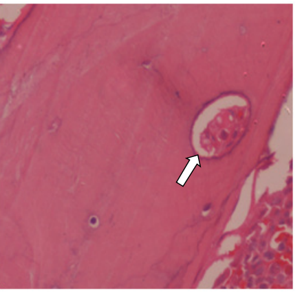
The purpose of this study was to develop standards for estimating age at death, using bone microstructure, that are applicable to a South Indian population. The sample consisted of 67 individuals (59 males and 8 females) of known age and sex. The sample was removed 5 cm lateral from the costo-chondral junction of the fourth riband slides were prepared according to standard histological methodology.
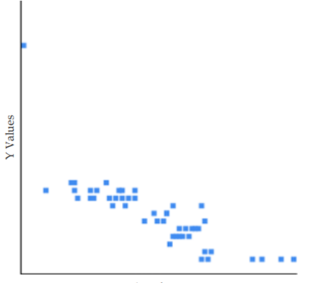
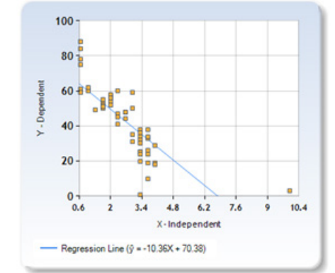
It was found that the number of Non-Haversian canals tend to reduce with age in a linear fashion which is also seen in the previous studies with coefficient of determination being 0.6703. Then the regression equation was calculated for estimating age using the number nonhaversian canal Y = –10.3637X + 70.37784 with standard error of estimate being ± 9.14 years.
References
- 1. Işcan MY, Loth SR, Wright RK. Age Estimation from the Rib by Phase Analysis: White Females. Journal of Forensic Sciences. 1985;30(3):853–863.
- 2. Işcan MY, Loth SR, Wright RK. Age Estimation from the Rib by Phase Analysis: White Males. Journal of Forensic Sciences. 1984;29(4):1094–1104.
- 3. Christensen AM, Crowder CM, Ousley SD, Houck MM. Error and its meaning in forensic science. Journal of Forensic Sciences. 2014;59(1):123–126.
- 4. Christensen AM, Passalacqua NV, Bartelink EJ. Age Estimation. In: Forensic Anthropology: Current Methods and Practice. Academic Press; 2014. p. 191–222.
- 5. Cappella A, Cummaudo M, Arrigoni E, Collini F, Cattaneo C. The issue of age estimation in a modern skeletal population: are even the more modern current aging methods satisfactory for the elderly? Journal of Forensic Sciences. 2017;62(1):12–17.
- 6. Stout SD, Paine RR. Brief communication: Histological age estimation using rib and clavicle. American Journal of Physical Anthropology. 1992;87(1):111–115.
- 7. Kerley ER. The microscopic determination of age in human bone. American Journal of Physical Anthropology. 1965;23(2):149–164.
- 8. Crowder CM, Heinrich J, Stout SD. Rib Histomorphometry for Adult Age Estimation. In: Crowder C, Stout S, editors. Bone Histology: An Anthropological Perspective. CRC Press; 2011. p. 235–248. (Note: Often cited as Humana Press 2012, but correctly CRC/Taylor & Francis).
- 9. Ericksen MF. Histologic estimation of age at death using the anterior cortex of the femur. American Journal of Physical Anthropology. 1991;84(2):171–179.
- 10. Maat GJR, Maes A, Aarents MJ, Nagelkerke NJD. Histological age prediction from the femur in a contemporary Dutch sample: The decrease of nonremodeled bone in the anterior cortex. Journal of Forensic Sciences. 2006;51(2):230–237.
Data Sharing Statement
There are no additional data available. All raw data and code are available upon request.
Funding
This research received no funding.
Author Contributions
Whether all authors contributed significantly to the work and approve its publication.
Ethics Declaration
This article does not involve any human or animal subjects, and therefore does not require ethics approval.
Acknowledgements
We would like to express our gratitude to the patients, their families, and all those who have contributed to this study.
Conflicts of Interest
The authors report no conflicts of interest in this work.
About this article
Cite this article
Chander UJK, Balasubramanian S, Srikrishnan S, et al. Can Vascular Channels in the Bone Determine the Age of A Human Being? Indian Journal of Forensic Medicine & Pathology. 2020;13(3):389–392.
Licence:
Attribution-Non-commercial 4.0 International (CC BY-NC 4.0)This license enables reusers to distribute, remix, adapt, and build upon the material in any medium or format for noncommercial purposes only, and only so long as attribution is given to the creator.
| Received | Accepted | Published |
|---|---|---|
| July 02, 2020 | July 20, 2020 | August 30, 2020 |
DOI: https://doi.org/10.21088/ijfmp.0974.3383.13320.4
Keywords
Non-Haversian canalRibAge estimationVascular channelsSearch for Similar Articles
Similar Articles
- Pesticide Contamination in Indian Agricultural and Residential Areas: A Compara...
- Forensic Age Estimation using CBCT-Derived Mandibular Morphometrics: A Comparat...
- A 2 Years Retrospective Study of the Spectrum of Poisoning in a Tertiary Care C...
- Knowledge and Attitude of MBBS Students Regarding Post Mortem Examination: A Cr...
- Sudden Death in a 7-year-old Child Due to Neoplasm: A Case Report
Article Level Metrics
Last UpdatedWednesday 08 July 2026, 07:39:10 (IST)
7723
Accesses
2
2137
00
Citations
NA
NA
NA
Download citation
Article Keywords
Keyword Highlighting
Highlight selected keywords in the article text.
Timeline
| Received | July 02, 2020 |
| Accepted | July 20, 2020 |
| Published | August 30, 2020 |
licence
Attribution-Non-commercial 4.0 International (CC BY-NC 4.0)
This license enables reusers to distribute, remix, adapt, and build upon the material in any medium or format for noncommercial purposes only, and only so long as attribution is given to the creator.