Full Text (PDF)
Indian Journal of Anatomy 7(5):p 502-507, Sep-Oct 2018. | DOI: http://dx.doi.org/10.21088/ija.2320.0022.7518.7
Original Article
A Study on the Morphometry of Occipital Condyles and Suboccipital Muscles in Human Dry Skulls and its Clinical Significance
Hema N., Associate Professor, Department of Anatomy, ESICÂMedical college & PGIMSR, Rajajinagar, Bengaluru, Karnataka 560010, India. , Swetha B.1 , Hema N.2
Author Information
Licence:
Indian Journal of Anatomy 7(5):p 502-507, Sep-Oct 2018. | DOI: http://dx.doi.org/10.21088/ija.2320.0022.7518.7
How Cite This Article:
Timeline
Received : N/A
Accepted : N/A
Published : N/A
Abstract
Introduction: The base of the human skull presents Occipital condyle which articulates with the atlas vertebra forming AtlantaOccipital joint. Keeping the head in inclined posture to engage in actions may play major role in architecture of facet. Maintenance of a particular posture demands more activity from muscles concerned. In this context the role of muscles in terms of traction to maintain a desired posture cannot be ruled out. More anatomical facts on the condyle facets will play an additional boon for the day to day surgeries.
Materials & Methods: One hundred adult South Indian skulls were examined using the scale, divider and the transparent centimeter reticule in the present study. The shape, size, the anteriorposterior diameter (length) and transverse diameter (width), surface area of occipital condyles on both sides were estimated. The location and number of extra facets were also noted. The surface area of suboccipital muscles were measured in search of the functional relation.
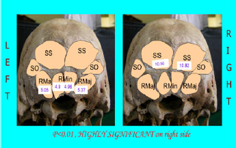
Results: Mean anteroposterior (length) and transverse diameter(width) of occipital condyles were 2.12cm & 1.1 cm on right side and 2.2 cm & 1.2 cm on left respectively. Mean Surface areas of the facet were 1.76 square centimeter (sq cm) on right and 1.9 sq cm on left which was significant. 22 extra facets were present maximally on the posteromedial aspect of the left occipital condyles. Its mean anteroposterior and transverse diameter were 0.52cm & 0.44 cm on right side and 0.45 cm &0.36 cm on left respectively. Mean surface areas (sq cm) of suboccipital muscles rectus capitis minor, rectus Capitis major and Obliquus Capitis Superior were 4.9, 5.05 and 10.56 on left side and 4.96,5.37 and 10.82 on right side respectively. Statistically the valued proved to be significant.
Conclusion: The present study provides anatomical knowledge on the measurements of the occipital condyles serves as a guide in various head and neck procedures for the Neurosurgeons and the Orthopaedicians. The incidence of the facets and the measurements of impressions of the suboccipital muscles on the occipital bone mentions the tilting posture of the humans on one side.
References
No records found.
About this article

Cite this article
Licence:
| Received | Accepted | Published |
|---|---|---|
| N/A | N/A | N/A |
DOI: http://dx.doi.org/10.21088/ija.2320.0022.7518.7
Keywords
Occipital Condyles; Extra Facets; Suboccipital Muscles.Search for Similar Articles
Similar Articles
- Supernumerary Pulmonary Vein: A Rare Anatomical Variation Observed During Cadave...
- Elongated Styloid Process: Anatomical Variations and Their Clinical Relevance: A...
- Bridging the Gap Between Dissection and Surgery: The Role of Soft Embalming in M...
- A Cadaveric Study of Retro Hepatic Segment of Inferior Vena Cava and Ostia Venae...
- Estimation of Stature from Forearm Length in Medical Undergraduate Students of a...
Article Level Metrics
Last UpdatedTuesday 22 September 2026, 05:43:58 (IST)
5059
Accesses
5
655
00
Citations
NA
NA
NA
Download citation
Article Keywords
Keyword Highlighting
Highlight selected keywords in the article text.
Timeline
| Received | N/A |
| Accepted | N/A |
| Published | N/A |