Full Text (PDF)
New Indian Journal of Surgery 15(1):p 27-31, January - March 2024. | DOI: https://dx.doi.org/10.21088/nijs.0976.4747.15124.4
Case Report
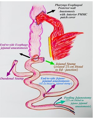
A Case Report of Corrosive Injury of Gastrointestinal Tract Managed with Pectoralis Major-Myocutaneous Patch Cover Over Pharyngo-Esophageal Anastomosis
Rajan Jagad, Arnab Chattopadhyay, Prashant Vanzare, Nidhi Shukla
Author Information
Licence:
Attribution-Non-commercial 4.0 International (CC BY-NC 4.0)This license enables reusers to distribute, remix, adapt, and build upon the material in any medium or format for noncommercial purposes only, and only so long as attribution is given to the creator.
New Indian Journal of Surgery 15(1):p 27-31, January - March 2024. | DOI: https://dx.doi.org/10.21088/nijs.0976.4747.15124.4
How Cite This Article:
Chattopadhyay A, Jagad R, Bhatt P, et al. A case report of corrosive injury of gastrointestinal tract managed with pectoralis major-myocutaneous patch cover over pharyngo-esophageal anastomosis. New Indian J Surg. 2024;15(3):27-31.Timeline
Received : December 11, 2023
Accepted : February 01, 2024
Published : March 24, 2024
Abstract
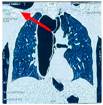
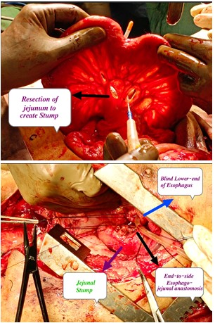
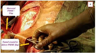
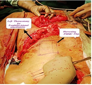
Corrosive ingestion remains one of the most devastating problems in developing countries that cripples patient’s body, mind and soul depriving them from leading a normal social life and living with a scar on their emotions, psyche and charisma. Appropriate management in the acute phase of corrosive injury reduces tissue damage and further morbidity and mortality. The aim of this case discussion is to evoke a light of hope for those patients of corrosive injury and a guide to clinicians worldwide for further better management and sharing our experience of surgical management of a case of such corrosive injury of gastrointestinal tract.
References
- 1. Jain R, Gupta S, Pasricha N, Faujdar M, Sharma M, Mishra P. ESCC with metastasis in the young age of caustic ingestion of shortest duration. J Gastrointest Cancer. 2010; 41:93-5.
- 2. Chirica M, Bonavina L, Kelly MD, Sarfati E, Cattan P. Caustic ingestion. Lancet. 2017; 389(10083):2041 2052. doi: 10.1016/S0140-6736(16)30313-0.
- 3. Ananthakrishnan N, Kalayarasan R, Kate V. Corrosive injury of esophagus and stomach. In: Mishra PK, editor. Textbook of Surgical Gastroenterology, 1st ed., New Delhi, India: Jaypee; 2016.
- 4. Lakshmi CP, Vijayahari R, Kate V, Anantha krishnan N. A hospital-based epidemiological study of corrosive alimentary injuries with particular reference to the Indian experience. Natl Med J India. 2013; 26(1):31–36.
- 5. Mowry JB, Spyker DA, Cantilena LR, Jr,, McMillan N, Ford M. 2013 Annual report of the american association of poison control centers’ national poison data system (NPDS): 31st annual report. ClinToxicol (Phila) 2014; 52(10):1032–1283.
- 6. Pankaja S, Valooran G, Das S, Suvasini L, Aravind R, Kate V, et al. Isolated full thickness jejunal necrosis following sulphuric acid cocktail ingestion—a clinical case report—isolated corrosive jejunal necrosis. Surg Sci. 2011; 2:338–340.
- 7. Osman M, Russell J, Shukla D, Moghadamfalahi M, Granger DN. Responses of the murine esophageal microcirculation to acute exposure to alkali, acid, or hypochlorite. J Pediatr Surg. 2008;43(9):1672 1678.
- 8. Genç A, Mutaf O. Esophageal motility changes in acute and late periods of caustic esophageal burns and their relation to prognosis in children. J Pediatr Surg. 2002;37:1526-8.
- 9. Tiryaki T, Livanelioğlu Z, Atayurt H. Early bougienage forrelief of stricture formation following caustic esophageal burns. Pediatr Surg Int. 2005;21:78-80.
Data Sharing Statement
There are no additional data available.
Funding
This research received no funding
Author Contributions
All authors contributed significantly to the work and approve its publication.
Ethics Declaration
This article does not involve any human or animal subjects, and therefore does not require ethics approval.
Acknowledgements
Information Not Provided
Conflicts of Interest
No conflicts of interest in this work.
About this article

Cite this article
Chattopadhyay A, Jagad R, Bhatt P, et al. A case report of corrosive injury of gastrointestinal tract managed with pectoralis major-myocutaneous patch cover over pharyngo-esophageal anastomosis. New Indian J Surg. 2024;15(3):27-31.
Licence:
Attribution-Non-commercial 4.0 International (CC BY-NC 4.0)This license enables reusers to distribute, remix, adapt, and build upon the material in any medium or format for noncommercial purposes only, and only so long as attribution is given to the creator.
| Received | Accepted | Published |
|---|---|---|
| December 11, 2023 | February 01, 2024 | March 24, 2024 |
DOI: https://dx.doi.org/10.21088/nijs.0976.4747.15124.4
Keywords
CorrosiveStrictureAnastomosisSearch for Similar Articles
Similar Articles
- Laparoscopy-Guided Bilateral Oblique Subcostal Transversus Abdominis Plane Bloc...
- Recognizing the Columnar Cell Subtype of Papillary Thyroid Carcinoma: A Case Rep...
- Randomized Controlled Study To Compare Intraperitoneal Plus Port-Site Instillat...
- Single Preoperative Dose Versus Conventional Multidose Antibiotic Prophylaxis In...
- To Compare the Dissection of Gallbladder by Ultrasonic Harmonic Scalpel Versus...
Article Level Metrics
Last UpdatedTuesday 29 September 2026, 15:46:08 (IST)
5241
Accesses
15
437
00
Citations
NA
NA
NA
Download citation
Article Keywords
Keyword Highlighting
Highlight selected keywords in the article text.
Timeline
| Received | December 11, 2023 |
| Accepted | February 01, 2024 |
| Published | March 24, 2024 |
licence
Attribution-Non-commercial 4.0 International (CC BY-NC 4.0)
This license enables reusers to distribute, remix, adapt, and build upon the material in any medium or format for noncommercial purposes only, and only so long as attribution is given to the creator.