Full Text (PDF)
New Indian Journal of Surgery 17(3):p 126-133, July - Sept. 2026. | DOI: 10.21088/nijs.0976.4747.17326.6
Original Article
Recognizing the Columnar Cell Subtype of Papillary Thyroid Carcinoma: A Case Report With Histopathologic and IHC Insights
Soumya B M, Rashmi P, Vardendra Kulkarni, R Gayathri, Smitha K Hegde
Author Information
Licence:
Attribution-Non-commercial 4.0 International (CC BY-NC 4.0)This license enables reusers to distribute, remix, adapt, and build upon the material in any medium or format for noncommercial purposes only, and only so long as attribution is given to the creator.
New Indian Journal of Surgery 17(3):p 126-133, July - Sept. 2026. | DOI: 10.21088/nijs.0976.4747.17326.6
How Cite This Article:
Soumya B M, Rashmi P, Vardendra Kulkarni. Recognizing The Columnar Cell Subtype of Papillary Thyroid Carcinoma: A Case Report With Histopathologic and IHC Insights. New Indian J Surg. 2026; 17(3): 126-133.Timeline
Received : June 15, 2026
Accepted : July 17, 2026
Published : September 30, 2026
Abstract
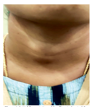
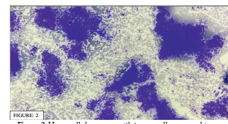
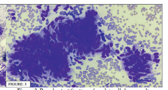
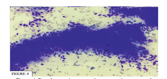
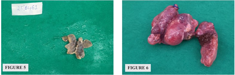
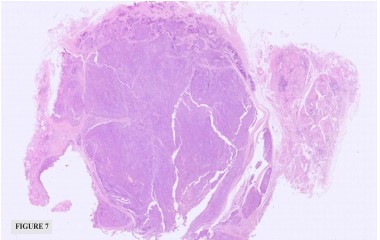
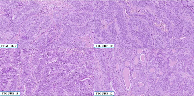
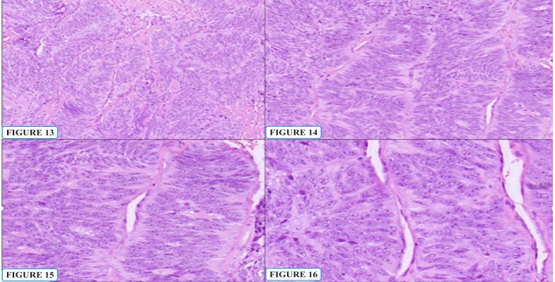
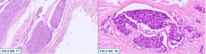
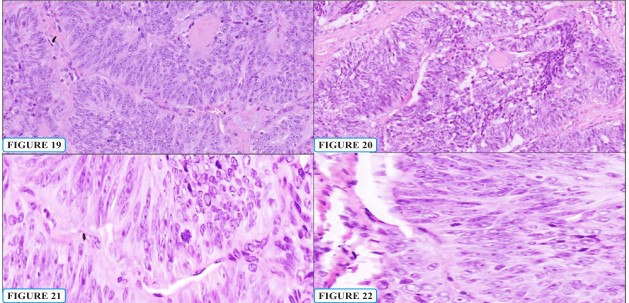
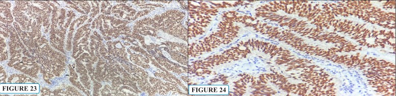
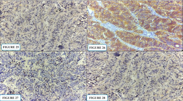
Columnar Cell subtype of Papillary Thyroid Carcinoma (CC-PTC) is an exceptionally rare histologic subtype, accounting for less than 0.5% of all papillary thyroid carcinomas worldwide.¹,² Despite its rarity, CC-PTC demonstrates aggressive biological behavior and is classified as a high-risk histologic subtype in the World Health Organization Classification of Tumors of Endocrine Organs, 5th Edition (2022)³, owing to its frequent association with capsular invasion, lymph vascular permeation, extrathyroidal extension, and distant metastasis. The tumor poses significant diagnostic challenges due to its distinctive morphology, particularly on cytology and limited biopsy material.4,6 Case Presentation: A 27-year-old woman presented with a one-year history of anterior neck swelling with recent onset throat pain. ltrasonography revealed a right thyroid lobe nodule categorized as TI-RADS 4/5. Fine-needle aspiration cytology showed highly cellular smears composed of tall and columnar epithelial cells arranged in papillary fragments and sheets, exhibiting nuclear elongation, hyperchromatic nuclei, marked pseudo stratification, and occasional nuclear pseudoinclusions, consistent with Bethesda Category VI. Total thyroidectomy revealed a 3-cm ill-defined, unencapsulated grey-white tumor. Histopathological and Immunohistochemical Findings: Histopathology demonstrated circumscribed tumor composed of complex papillary and trabecular architecture lined by pseudostratified columnar cells with elongated hyperchromatic nuclei, conspicuous eosinophilic nucleoli, amphophilic cytoplasm, increased mitotic activity, and extensive lymphovascular invasion. Immunohistochemistry confirmed thyroid follicular epithelial origin with tumor cell nuclear positivity for TTF-1 and retained membranous β-catenin expression, while CDX2 was negative. A high Ki-67 labelling index supported the aggressive proliferative nature of the tumor. Conclusion: This case highlights the importance of recognizing the distinctive cytomorphologic, histopathologic and immunohistochemical features of CC-PTC. Accurate diagnosis requires a multimodal approach integrating clinical findings, cytology, histopathology, and immunohistochemistry, particularly in young patients where aggressive behavior may be unsuspected.
References
- 1. Lloyd RV, Osamura RY, Klöppel G, Rosai J, Editors. WHO Classification Of Tumours Of Endocrine Organs. 5th Ed. Lyon: International Agency For Research On Cancer; 2022.
- 2. Asioli S, Erickson LA, Sebo TJ, Zhang J, Jin L, Thompson GB, et al. Papillary thyroid carcinoma with prominent columnar cell features: a clinicopathologic study of 17 cases. Am J Surg Pathol. 2010;34(5):662–669. doi:10.1097/PAS.0b013e3181d96c57.
- 3. Mete O, Asa SL. Pathological definition and clinical significance of aggressive variants of papillary thyroid carcinoma. J Clin Endocrinol Metab. 2011;96(6):1806–1814. doi:10.1210/ jc.2010-2877.
- 4. Wenig BM. Aggressive variants of papillary thyroid carcinoma. Mod Pathol. 2011;24(Suppl 2):S18–S27. doi:10.1038/modpathol.2010.131.
- 5. LiVolsi VA. Papillary thyroid carcinoma: an update. Mod Pathol. 2011;24(Suppl 2):S1–S9. doi:10.1038/modpathol.2010.129.
- 6. Chen JH, Faquin WC. Diagnosis of papillary thyroid carcinoma variants on fine-needle aspiration biopsy. Cancer Cytopathol. 2012;120(6):407–414. doi:10.1002/cncy.21225.
- 7. Baloch ZW, LiVolsi VA. Fine-needle aspiration of thyroid nodules: past, present, and future. Endocr Pract. 2004;10(3):234–241. doi:10.4158/ EP.10.3.234.
- 8. Bongiovanni M, Spitale A, Faquin WC, Mazzucchelli L, Baloch ZW. The Bethesda System for Reporting Thyroid Cytopathology: a meta-analysis. Acta Cytol. 2012;56(4):333– 339. doi:10.1159/000339959.
- 9. Ordonez NG. Value of thyroid transcription factor-1 immunostaining in tumor diagnosis: a review and update. Appl Immunohistochem Mol Morphol. 2012;20(5):429–444. doi:10.1097/ PAI.0b013e318254056b.
- 10. Xu B, Ghossein R. Evolution of the histologic classification of thyroid neoplasms and its impact on clinical management. Eur J Surg Oncol. 2018;44(3):338–347. doi:10.1016/j. ejso.2017.11.016.
- 11. Rivera M, Tuttle RM, Patel S, Shaha A, Shah JP, Ghossein RA. Encapsulated papillary thyroid carcinoma: a clinicopathologic study of 106 cases. Hum Pathol. 2009;40(12):1708–1713. doi:10.1016/j.humpath.2009.04.007.
- 12. Tallini G, Tuttle RM, Ghossein RA. The history of the follicular variant of papillary thyroid carcinoma. J Clin Endocrinol Metab. 2017;102(1):15–22. doi:10.1210/jc.2016-2970.
- 13. Evans HL. Columnar-cell carcinoma of the thyroid. A report of nine cases of a distinctive type of thyroid carcinoma. Am J Surg Pathol. 1986;10(11):737–749.
Data Sharing Statement
There are no additional data available. All raw data and code are available upon request.
Funding
This research received no funding.
Author Contributions
All authors contributed significantly to the work and approve its publication.
Ethics Declaration
This article does not involve any human or animal subjects, and therefore does not require ethics approval.
Acknowledgements
We would like to express our gratitude to the patients, their families, and all those who have contributed to this study.
Conflicts of Interest
No conflicts of interest in this work.
About this article

Cite this article
Soumya B M, Rashmi P, Vardendra Kulkarni. Recognizing The Columnar Cell Subtype of Papillary Thyroid Carcinoma: A Case Report With Histopathologic and IHC Insights. New Indian J Surg. 2026; 17(3): 126-133.
Licence:
Attribution-Non-commercial 4.0 International (CC BY-NC 4.0)This license enables reusers to distribute, remix, adapt, and build upon the material in any medium or format for noncommercial purposes only, and only so long as attribution is given to the creator.
| Received | Accepted | Published |
|---|---|---|
| June 15, 2026 | July 17, 2026 | September 30, 2026 |
DOI: 10.21088/nijs.0976.4747.17326.6
Keywords
Papillary Thyroid CarcinomaColumnar Cell VariantFine-Needle Aspiration CytologyImmunohistochemistryKi-67B-CateninCDX2Search for Similar Articles
Similar Articles
- Laparoscopic Repair of Large Recurrent Bochdalek Hernia: Case Report with Review...
- A Retrospective Evaluation of Superior Efficacy in Irrigation and Flushing of Sa...
- Implementation of Enhanced Recovery After Surgery (ERAS) Protocols in Elective C...
- Accuracy of TIRADS Classification in the Risk Stratification of Thyroid Swelling...
- Case Series Analysis of Medical Expulsive Therapy in Ureteric Calculi
Article Level Metrics
Last UpdatedSunday 18 October 2026, 07:46:53 (IST)
7249
Accesses
22
787
00
Citations
NA
NA
NA
Download citation
Article Keywords
Keyword Highlighting
Highlight selected keywords in the article text.
Timeline
| Received | June 15, 2026 |
| Accepted | July 17, 2026 |
| Published | September 30, 2026 |
licence
Attribution-Non-commercial 4.0 International (CC BY-NC 4.0)
This license enables reusers to distribute, remix, adapt, and build upon the material in any medium or format for noncommercial purposes only, and only so long as attribution is given to the creator.