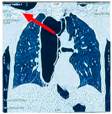
CT-scan with Red arrow showing Dilated megaesophagus, slightly deviated to right mediastinum with blind lower-end.
Description: No description available.
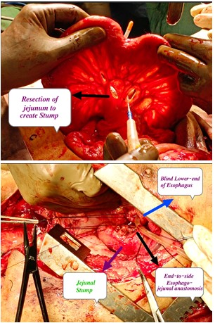
Showing image A with Resection of jejunum to create stump, Image B with end-to-side esophago-jejunal anastomosis.
Description: No description available.
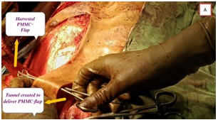
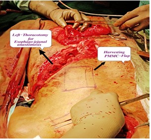
Showing Image A with creation of tunnel in left cervical region to deliver newly harvested PMMC-Flap, Image B Harvesting left sided PMMC-Flap
Description: No description available.
Showing Image A with creation of tunnel in left cervical region to deliver newly harvested PMMC-Flap, Image B Harvesting left sided PMMC-Flap
Description: No description available.
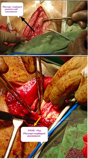
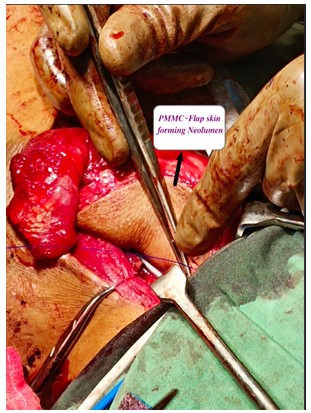
Showing Image A with Pharyngo-esophageal posterior wall anastomosis, Image B with PMMC-Flap Pharyngo-esophageal anastomosis, Image C with Neolumen formed by inverted skin of PMMC-Flap.
Description: No description available.
Showing Image A with Pharyngo-esophageal posterior wall anastomosis, Image B with PMMC-Flap Pharyngo-esophageal anastomosis, Image C with Neolumen formed by inverted skin of PMMC-Flap.
Description: No description available.
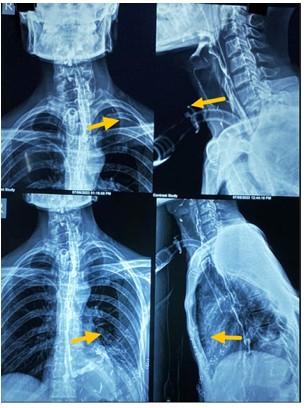
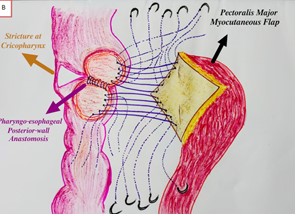
showing image A with Barium swallow showing easy flow of contrast through upper pharyngo-esophageal PMMC Flap anastomosis and lower esophago-jejunal anastomosis without any leak of contrast, Image B with pictorial presentation of pharyngo-esophageal PMMC Flap- anastomosis.
Description: No description available.
showing image A with Barium swallow showing easy flow of contrast through upper pharyngo-esophageal PMMC Flap anastomosis and lower esophago-jejunal anastomosis without any leak of contrast, Image B with pictorial presentation of pharyngo-esophageal PMMC Flap- anastomosis.
Description: No description available.
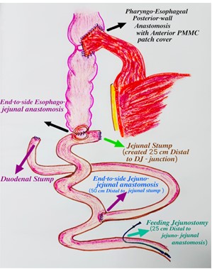
Showing Image with pictorial presentation of various anastomosis that are performed