Full Text (PDF)
Indian Journal of Anatomy 13(2):p 75-77, April - June 2024. | DOI: https://doi.org/10.21088/ija.2320.0022.13224.5
Case Report
A Case of Double Profunda Femoris Artery
Swati Sharma, Ratesh Kumar Munjal, Biddyabati Patowari
Author Information
Licence:
Attribution-Non-commercial 4.0 International (CC BY-NC 4.0)This license enables reusers to distribute, remix, adapt, and build upon the material in any medium or format for noncommercial purposes only, and only so long as attribution is given to the creator.
Indian Journal of Anatomy 13(2):p 75-77, April - June 2024. | DOI: https://doi.org/10.21088/ija.2320.0022.13224.5
How Cite This Article:
8. Sharma S, Munjal RK, Patowari B. A Case of Double Profunda Femoris Artery. Indian J Anat. 2024; 13(2): 75-77.Timeline
Received : May 12, 2024
Accepted : August 14, 2024
Published : September 30, 2024
Abstract
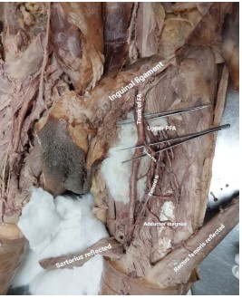
During a routine dissection of an adult male cadaver for academic purpose two Profunda Femoris Arteries (PFA) were seen arising from Femoral Artery (FA). Both were following an expected course. Double PFA is an extremely rare anatomical variation, only a few such cases are reported. The two PFA, upper and lower were found to be arising 2 cm and 5 cm from Mid Inguinal Point (MIP) respectively. A thorough knowledge of variations of PFA is of great importance to avoid complications during surgical procedures. The variations in PFA and its branches are very common and largely associated with one another. The PFA has an important role in collateral blood flow in atherosclerotic occlusive disease. The common femoral artery is frequently accessed by radiologists and surgeons and is easily accessible to catheterization. Knowledge of the Anatomy and variations in PFA is very important for surgeons to avoid any iatrogenic injury to the patient.
References
- 1. Dutta A. Essentials of Human Anatomy: Superior & Inferior Extremities. 5th ed. Kolkata: Current Books International; 2017. p. 144.
- 2. Tsovcalas G, Panagovli E, Fiska A, et al. A rare double profunda femoris artery in a female cadaver. Abc. 2018; p. 212-213.
- 3. Lakshmi V, Seshagyan S, Chitra S. Anatomical variation of profunda femoris artery. Int J Curr Med Appl Sci. 2016; 10(1): 1-3.
- 4. George A, Santhakumary M. A cadaveric study on the variation of the profunda femoris artery in South India. Asian J Med Sci. 2021; 12(2): 86-90.
- 5. Manpappa T, Prasanna L. Anatomical variation of profunda femoris artery and its branches: A cadaveric study in South Indian population. Indian J Surg. 2014; 76(4): 288-292.
- 6. Waghmode S, Syed S. Anatomical variation of profunda femoris artery in Indian population. Ann Int Med Den Res. 2022; 8(1): 180-184.
- 7. Quazi E, Wilting J, Patel N, et al. Arteries of lower limb: Embryology, variation and clinical significance. Can Assoc Radiol J. 2022; 73(1): 259-270.
Data Sharing Statement
There are no additional data available. All raw data and code are available upon request.
Funding
This research received no funding.
Author Contributions
Whether all authors contributed significantly to the work and approve its publication.
Ethics Declaration
This article does not involve any human or animal subjects, and therefore does not require ethics approval.
Acknowledgements
We would like to express our gratitude to the patients, their families, and all those who have contributed to this study.
Conflicts of Interest
The authors report no conflicts of interest in this work.
About this article

Cite this article
8. Sharma S, Munjal RK, Patowari B. A Case of Double Profunda Femoris Artery. Indian J Anat. 2024; 13(2): 75-77.
Licence:
Attribution-Non-commercial 4.0 International (CC BY-NC 4.0)This license enables reusers to distribute, remix, adapt, and build upon the material in any medium or format for noncommercial purposes only, and only so long as attribution is given to the creator.
| Received | Accepted | Published |
|---|---|---|
| May 12, 2024 | August 14, 2024 | September 30, 2024 |
DOI: https://doi.org/10.21088/ija.2320.0022.13224.5
Keywords
Anatomical VariationLower limbTwo Profunda Femoris ArterySearch for Similar Articles
Similar Articles
- Supernumerary Pulmonary Vein: A Rare Anatomical Variation Observed During Cadave...
- Elongated Styloid Process: Anatomical Variations and Their Clinical Relevance: A...
- Bridging the Gap Between Dissection and Surgery: The Role of Soft Embalming in M...
- A Cadaveric Study of Retro Hepatic Segment of Inferior Vena Cava and Ostia Venae...
- Estimation of Stature from Forearm Length in Medical Undergraduate Students of a...
Article Level Metrics
Last UpdatedSunday 20 September 2026, 22:34:42 (IST)
5038
Accesses
10
601
00
Citations
NA
NA
NA
Download citation
Article Keywords
Keyword Highlighting
Highlight selected keywords in the article text.
Timeline
| Received | May 12, 2024 |
| Accepted | August 14, 2024 |
| Published | September 30, 2024 |
licence
Attribution-Non-commercial 4.0 International (CC BY-NC 4.0)
This license enables reusers to distribute, remix, adapt, and build upon the material in any medium or format for noncommercial purposes only, and only so long as attribution is given to the creator.