Full Text (PDF)
Indian Journal of Pathology: Research and Practice 11(2):p 53-60, April-June 2022. | DOI: https://doi.org/10.21088/ijprp.2278.148X.11222.5
Original Article
Use of C3d and C4d Immunohistological Markers to Differentiate Various Inflammatory Dermatosis
Parimal Ravindra Gode, Sharada Raju Rane
Author Information
Licence:
Attribution-Non-commercial 4.0 International (CC BY-NC 4.0)This license
enables reusers to distribute, remix, adapt, and build upon the material in any
medium or format for noncommercial purposes only, and only so long as
attribution is given to the creator.
Indian Journal of Pathology: Research and Practice 11(2):p 53-60, April-June 2022. | DOI: https://doi.org/10.21088/ijprp.2278.148X.11222.5
How Cite This Article:
Parimal Ravindra Gode, Sharada Raju Rane/Use of C3d and C4d Immunohistological Markers to Differentiate Various Inflammatory Dermatosis”/Indian Journal of Pathology: Research and Practice 2022;11(2) 53–60.Timeline
Received : January 17, 2022
Accepted : February 25, 2022
Published : June 25, 2022
Abstract
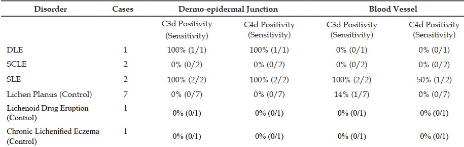
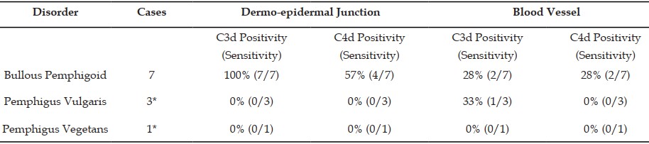
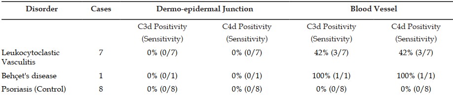
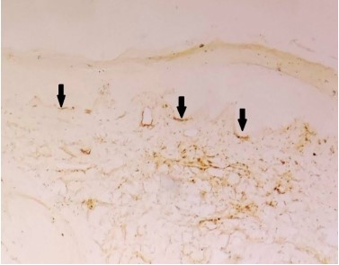
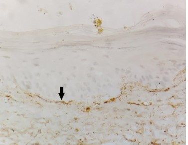
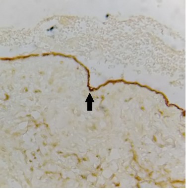
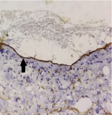
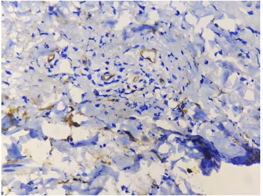
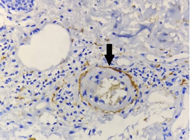
Introduction: The application of C3d and C4d as a diagnostic adjunct in the cases of inflammatory skin disease has been rarely reported in the literature. The purpose of our study is to define the significance of C3d and C4d deposition by an immunohistochemistry (IHC) on paraffin-embedded, formalin-fixed tissue as a correlative diagnostic adjunct in the assessment of inflammatory skin disease. Objective: To evaluate use of C3d and C4d by immunohistochemistry (IHC) on formalin-fixed tissue as diagnostic adjunct in the various inflammatory skin diseases. Method: 1) Diagnosed cases of inflammatory skin diseases on histology were selected for the study. 2) H&E slides were studied for morphological assessment. 3) IHC makers for C3d and C4d were performed on the paraffin blocks using C3d and C4d- antibody based detection kits. Result: 100% cases of DLE & SLE were positive for deposition of C3d & C4d at dermo-epidermal junction. 100% cases of Bullous Pemphigoid & 57% of cases of Bullous Pemphigoid were positive for C3d and C4d deposition respectively at dermo-epidermal junction. 100% cases of Behçet's disease & 42% cases ofLeukocytoclastic Vasculitis were positive for deposition of C3d & C4d in blood vessel. Conclusion: The C3d and C4d assays define an important diagnostic adjunct in the evaluation of collagen vascular disease, cutaneous vascular disease, and autoimmune vesiculobullous disease. The assays may prompt further DIF testing or, in some instances, may even define a reasonable substitute for DIF and/or add to the morphologic assessment of a biopsy specimen submitted for routine light microscopic assessment.
References
- 1. Giang J, Seelen MAJ, Doorn MBA Van, Rissmann R. Complement Activation in inflammatory Skin Diseases. 2018;9(April):1–17.
- 2. Troxell M, Weintraub L, Higgins J, Kambham N. Comparison of C4d immunostaining methods in renal allograft biopsies. Clin J Am Soc Nephrol. 2006;1:583–91.
- 3. Rodriguez ER, Skojec D V., Tan CD, Zachary AA, Kasper EK, Conte J V., et al. Antibody-mediated rejection in human cardiac allografts: Evaluation of immunoglobulins and complement activation products C4d and C3d as markers. Am J Transplant. 2005;5(11):2778–85.
- 4. Haas M, Rahman M, Racusen L, Kraus E, Bagnasco S, Segev D. C4d and C3d staining in biopsies of ABO- and HLA-incompatible renal allografts: correlation with histologic findings. Am J Transpl. 2006;6:1829–40.
- 5. Magro CM, Dyrsen ME. The use of C3d and C4d immunohistochemistry on formalin-fixed tissue as a diagnostic adjunct in the assessment of inflammatory skin disease. J Am Acad Dermatol [Internet]. 2008;59(5):822–33. Available from: http://dx.doi.org/10.1016/j.jaad.2008.06.022
- 6. Pfaltz K, Mertz K, Rose C, Scheidegger P, Pfaltz M, Kempf W. C3d immunohistochemistry on formalinfixed tissue is a valuable tool in the diagnosis of bullous pemphigoid of the skin. J Cutan Pathol. 2010;37(6):654–8.
- 7. Chandler W, Zone J, Florell S. C4d immunohistochemical stain is a sensitive method to confirm immunoreactant deposition in formalinfixed paraffin-embedded tissue in bullous pemphigoid. J Cutan Pathol. 2009;36(6):655–9.
- 8. Schrager M, Rothfield N. Pathways of complement activation in chronic discoid lupus: serologic and immunofluorescence studies. Arthritis Rheum. 1977;(20):637–45.
- 9. Smith C, Marino C, Rothfield N. The clinical utility of the lupus band test. Arthritis Rheum. 1984;(27):382–7.
- 10. de Sousa Pinto JM, Sacramento Marques M, Estanislau Correia T. Lichen planus and leukocytoclastic vasculitis induced by interferon alpha-2b in a subject with HCV-related chronic active hepatitis. J Eur Acad Dermatology Venereol. 2003;17(2):193–5.
Data Sharing Statement
There are no additional data available. All raw data and code are available upon request.
Funding
This research received no funding.
Author Contributions
All authors contributed significantly to the work and approve its publication.
Ethics Declaration
This article does not involve any human or animal subjects, and therefore does not require ethics approval.
Acknowledgements
We would like to express our gratitude to the patients, their families, and all those who have contributed to this study.
Conflicts of Interest
No conflicts of interest in this work.
About this article

Cite this article
Parimal Ravindra Gode, Sharada Raju Rane/Use of C3d and C4d Immunohistological Markers to Differentiate Various Inflammatory Dermatosis”/Indian Journal of Pathology: Research and Practice 2022;11(2) 53–60.
Licence:
Attribution-Non-commercial 4.0 International (CC BY-NC 4.0)This license
enables reusers to distribute, remix, adapt, and build upon the material in any
medium or format for noncommercial purposes only, and only so long as
attribution is given to the creator.
| Received | Accepted | Published |
|---|---|---|
| January 17, 2022 | February 25, 2022 | June 25, 2022 |
DOI: https://doi.org/10.21088/ijprp.2278.148X.11222.5
Keywords
Search for Similar Articles
Similar Articles
- Clinico-Pathological and Immunohistochemical Study of Primary CNS Tumors in a Te...
- The Role of SGLT2 Inhibitors in Cardiovascular Protection Beyond Diabetes: A Co...
- Epidermal Inclusion Cyst Presenting as 4th Ventricle Space Occupying Lesion: A R...
- Silent Retroperitoneal Seminoma Arising from an Occult Undescended Testis: Diagn...
- Scapular Metastasis of Oral Squamous Cell Carcinoma: A Case Report of an Unusual...
Article Level Metrics
Last UpdatedSunday 20 September 2026, 22:44:03 (IST)
3662
Accesses
11
541
00
Citations
NA
NA
NA
Download citation
Article Keywords
Keyword Highlighting
Highlight selected keywords in the article text.
Timeline
| Received | January 17, 2022 |
| Accepted | February 25, 2022 |
| Published | June 25, 2022 |
licence
Attribution-Non-commercial 4.0 International (CC BY-NC 4.0)
This license
enables reusers to distribute, remix, adapt, and build upon the material in any
medium or format for noncommercial purposes only, and only so long as
attribution is given to the creator.