Full Text (PDF)
Indian Journal of Forensic Medicine and Pathology 19(3):p 381-384, July -Sept. 2026. | DOI: https://doi.org/10.21088/ijfmp.0974.3383.19326.14
Case Series
Sudden Death: The Unseen Danger of Tuberculosis
Ganesh Rajahan, Syed Affridh, Venkatesan. M., Sneha S., Priyadarshee Pradhan
Author Information
Licence:
Attribution-Non-commercial 4.0 International (CC BY-NC 4.0)This license enables reusers to distribute, remix, adapt, and build upon the material in any medium or format for noncommercial purposes only, and only so long as attribution is given to the creator.
Indian Journal of Forensic Medicine and Pathology 19(3):p 381-384, July -Sept. 2026. | DOI: https://doi.org/10.21088/ijfmp.0974.3383.19326.14
How Cite This Article:
Syed Affridh, Ganesh Rajahan, Venkatesan M. et al. Sudden Death: The Unseen Danger of Tuberculosis. Indian J Forensic Med Pathol. 2026; 19(3): 381-384Timeline
Received : March 17, 2026
Accepted : May 26, 2026
Published : September 30, 2026
Abstract
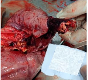
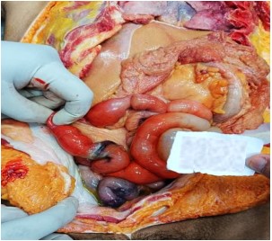
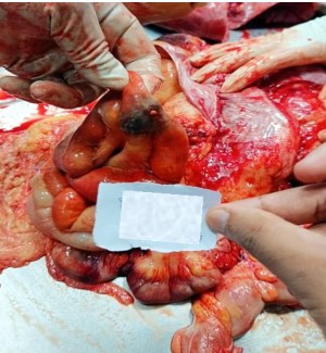
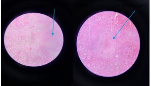
Tuberculosis still remains a major global health problem, especially in developing countries. Death and prevalence of tuberculosis have been reported worldwide. Tuberculosis usually presents primarily as lung disease with dissemination to the intestines increasing its morbidity. But the challenging part is its vague symptoms. Sudden death following intestinal TB is rare. A case involving a 24-year-old woman was received in unconscious state at the emergency department with complaints of severe stomach pain and vomiting. Despite resuscitative measures, she was declared dead. The postmortem examination revealed a white, cheeselike area at the apex of her right lung. Thickened dark tissue noted over the ileocecal junction. Histopathology confirmed tuberculosis in both lungs and intestines. Intestinal obstruction was noted at the site of TB involvement. This case highlights the importance of recognizing severe forms of intestinal tuberculosis this case underlines the vital role of precise medicolegal autopsy and complete histopathological examination in identifying hidden causes of sudden death.
References
- 1. World Health Organization. Tuberculosis. WHO, 2024.
- 2. Stat Pearls Publishing. Digestive Tract Tuberculosis.
- 3. Horvath K.D., Whelan R.L. Intestinal tuberculos is: return of an old disease. Am J Gastroenterol. 1998; 93: 692–696.
- 4. Sharma S.K., Mohan A. Extrapulmonary tuberculosis. Indian J Med Res. 2004.
- 5. Ma J.Y., et al. Intestinal tuberculosis and Crohn’s disease: diagnostic challenges. World J Gastroenterol. 2016; 22: 155–161.
- 6. Ha H.K., et al. Abdominal tuberculosis: radiologic features and differential diagnosis. Radio graphics. 1999; 19: 32–38.
- 7. Weedon D. Granulomasinderma to pathology. Dermatopathology. 2021.
- 8. Na J.Y., et al. Suddenun expected death caused by intestinal tuberculosis. Medicine (Baltimore). 2015.
- 9. Durney B.A., et al. Gastrointestinal tuberculosis review. Clin Gastroenterol. 2018.
- 10. Aminullah A., et al. Intestinal tuberculosis presenting with obstruction. J ClinDiagn Res. 2017; 11: 52–54.
- 11. Tobin D.M., Khatri S. Pathogenesis of intestinal tuberculosis. Lancet Infect Dis. 2025.
- 12. Huang Y., et al. Intestinal tuber culosis present in gas bowel obstruction. BMC Gastroenterol. 2024; 24: 248–256.
- 13. Chinen K., Ito M. Fatal intestinal tuberculosis: autopsy report. Pathol Int. 2019.
- 14. Sharma M.P., Bhatia V. Abdominal tuberculos is. Indian J Med Res. 2004.
- 15. Ahuja A., et al. Histopathological spectrum of tuber culosis. Cureus. 2024.
- 16. WuXR, et al. Intestinal tuberculosis causes obstruction and perforation. World J Gastroenterol. 2015.
- 17. Kumar B., et al. Sudden death related to tuberculous myocarditis: Autopsy report. Forensic Sci Med Pathol. 2025
Data Sharing Statement
There are no additional data available. All raw data and code are available upon request.
Funding
This research received no funding.
Author Contributions
All authors contributed significantly to the work and approve its publication.
Ethics Declaration
This article does not involve any human or animal subjects, and therefore does not require ethics approval.
Acknowledgements
We would like to express our gratitude to the patients, their families, and all those who have contributed to this study.
Conflicts of Interest
No conflicts of interest in this work.
About this article

Cite this article
Syed Affridh, Ganesh Rajahan, Venkatesan M. et al. Sudden Death: The Unseen Danger of Tuberculosis. Indian J Forensic Med Pathol. 2026; 19(3): 381-384
Licence:
Attribution-Non-commercial 4.0 International (CC BY-NC 4.0)This license enables reusers to distribute, remix, adapt, and build upon the material in any medium or format for noncommercial purposes only, and only so long as attribution is given to the creator.
| Received | Accepted | Published |
|---|---|---|
| March 17, 2026 | May 26, 2026 | September 30, 2026 |
DOI: https://doi.org/10.21088/ijfmp.0974.3383.19326.14
Keywords
TuberculosisSudden deathIntestinal TBIntestinal obstructionSearch for Similar Articles
Similar Articles
- Histopathological Spectrum of Autopsy Cases: A One Year Retrospective Observatio...
- Forensic Differentiation of Manner of Death in Railway Fatalities: A FourCase Se...
- Understanding assault Patterns: A Cross-sectional study of Cases at Chri, Kelamb...
- Assessing Response Awareness to a Snake Bite in Population of Pune Region
- A Study on: Child Sexual Abuse among the Urban Population: Knowledge, Experience...
Article Level Metrics
Last UpdatedTuesday 15 September 2026, 12:08:42 (IST)
13302
Accesses
9
2854
00
Citations
NA
NA
NA
Download citation
Article Keywords
Keyword Highlighting
Highlight selected keywords in the article text.
Timeline
| Received | March 17, 2026 |
| Accepted | May 26, 2026 |
| Published | September 30, 2026 |
licence
Attribution-Non-commercial 4.0 International (CC BY-NC 4.0)
This license enables reusers to distribute, remix, adapt, and build upon the material in any medium or format for noncommercial purposes only, and only so long as attribution is given to the creator.