Full Text (PDF)
Indian Journal of Anatomy 9(4):p 243-250, October-December 2020. | DOI: http://dx.doi.org/10.21088/ija.2320.0022.9420.2
Original Article
Morpho–Metrical and Stereological Analysis of Human Fetal Kidney During Mid-Gestation Period Ranging from 12th to 35th Weeks to Study Growth Pattern of Kidney in Qualitative and Quantitative Aspects
Kirti Sudhakarraoo Solanke , Kirti Sudhakarrao Solanke1 , Rajan Bhatnagar2 , Boruah Dibyajyoti3 , Tandon Aseem4
Author Information
Licence:
Indian Journal of Anatomy 9(4):p 243-250, October-December 2020. | DOI: http://dx.doi.org/10.21088/ija.2320.0022.9420.2
How Cite This Article:
Kirti Sudhakarrao Solanke, Rajan Bhatnagar, Boruah Dibyajyoti, et al. Morpho–Metrical and Stereological Analysis of Human Fetal Kidney During Mid-Gestation Period Ranging from 12th to 35th Weeks to Study Growth Pattern of Kidney in Qualitative and
Quantitative Aspects. Indian J Anat. 2020;9(4):243–250.
Timeline
Received : N/A
Accepted : N/A
Published : N/A
Abstract
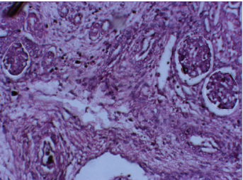
Morpho–metrical and stereological analysis of human fetal kidney during mid-gestation period ranging from 12th to 35th weeks of gestation to study growth pattern of kidney in qualitative and quantitative aspects. Aims and objective: Carry out Morpho-metrical Analysis of human fetal kidney during midgestation period ranging from 12th to 35th week and evaluating following parameters: a. Physical dimensions and weight of each kidney. b. Glomerular area for each kidney at different gestational ages. c. The numerical density of glomeruli in the cortex of each kidney at different gestational ages. d. Volume of glomeruli in the 3 different zones of each kidney at different gestational ages. Methods: For study purpose we divided 31 aborted fetuses into six groups according to gestational age. After measuring external parameters such as CRL of fetuses; they were injected with 10% formalin and fixed in 10% formalin for 3 days. Kidneys were procured; physical dimensions, volume and weight of kidney were measured. After tissue processing, blocks were cut; sections were mounted and H&E stained. Using software analyzer density, area, volume of glomeruli at three different zones i.e. cortex, juxtramedullary and medullary regions were calculated. Glomerular area and volume of cortical, juxtamedullary and medullary region for each kidney at different gestational ages was estimated by measuring the area and volume of 100 glomeruli per fetal kidney under 20X objective lens of image analyzer. Comparison was done for three different zones.
References
No records found.
About this article

Cite this article
Kirti Sudhakarrao Solanke, Rajan Bhatnagar, Boruah Dibyajyoti, et al. Morpho–Metrical and Stereological Analysis of Human Fetal Kidney During Mid-Gestation Period Ranging from 12th to 35th Weeks to Study Growth Pattern of Kidney in Qualitative and
Quantitative Aspects. Indian J Anat. 2020;9(4):243–250.
Licence:
| Received | Accepted | Published |
|---|---|---|
| N/A | N/A | N/A |
DOI: http://dx.doi.org/10.21088/ija.2320.0022.9420.2
Keywords
Kidney; Glomeruli; Density; Steriology; Volume; Area; Cortex; MedullaSearch for Similar Articles
Similar Articles
- Supernumerary Pulmonary Vein: A Rare Anatomical Variation Observed During Cadave...
- Elongated Styloid Process: Anatomical Variations and Their Clinical Relevance: A...
- Bridging the Gap Between Dissection and Surgery: The Role of Soft Embalming in M...
- A Cadaveric Study of Retro Hepatic Segment of Inferior Vena Cava and Ostia Venae...
- Estimation of Stature from Forearm Length in Medical Undergraduate Students of a...
Article Level Metrics
Last UpdatedMonday 21 September 2026, 23:42:45 (IST)
5056
Accesses
6
655
00
Citations
NA
NA
NA
Download citation
Article Keywords
Keyword Highlighting
Highlight selected keywords in the article text.
Timeline
| Received | N/A |
| Accepted | N/A |
| Published | N/A |